Volume 21, Number 4—April 2015
Dispatch
Candidate New Rotavirus Species in Sheltered Dogs, Hungary
Eszter Mihalov-Kovács, Ákos Gellért, Szilvia Marton, Szilvia L. Farkas, Enikő Fehér, Miklós Oldal, Ferenc Jakab, Vito Martella, and Krisztián Bányai
Figure 1
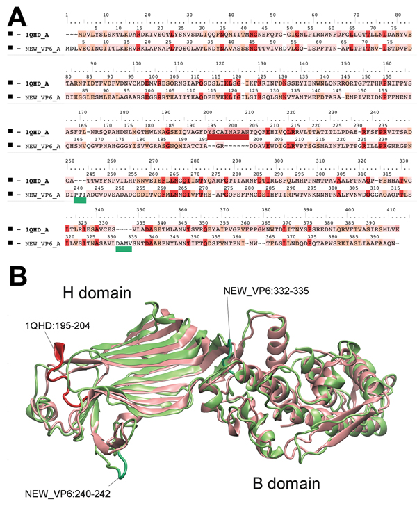
Figure 1. Structure comparison of rotavirus viral protein (VP) 6 proteins. A) Structure-based amino acid sequence alignment of the novel canine rotavirus VP6 protein and the template bovine rotavirus A VP6 protein. The background of the sequence alignments reflects the homology levels of the 2 VP6 sequences. Red, identical amino acid; orange, similar amino acid; pink, different amino acid). The main structural differences are indicated by dark red and menthol green on the sequence alignment and on the superimposed VP6 structures (B). Cartoon presentation of the homologous VP6 proteins: pink, rotavirus A; green, rotavirus I. Further information is available in the Technical Appendix.
Page created: March 17, 2015
Page updated: March 17, 2015
Page reviewed: March 17, 2015
The conclusions, findings, and opinions expressed by authors contributing to this journal do not necessarily reflect the official position of the U.S. Department of Health and Human Services, the Public Health Service, the Centers for Disease Control and Prevention, or the authors' affiliated institutions. Use of trade names is for identification only and does not imply endorsement by any of the groups named above.