Volume 23, Number 2—February 2017
Research
Highly Pathogenic Influenza A(H5Nx) Viruses with Altered H5 Receptor-Binding Specificity
Figure 2
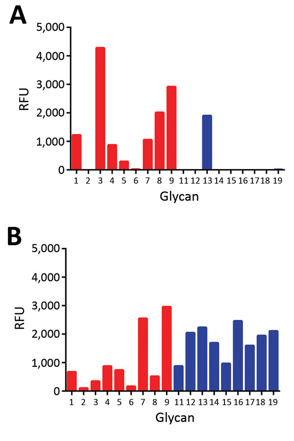
Figure 2. Glycan array analysis of recombinant H5 proteins of influenza A viruses. A) Wild-type H5N12.3.4 (KS) and B) H5N8 (QR) H5 proteins were applied to the glycan array precomplexed with StrepMAB-classic (IBA GmbH, Göttingen, Germany) and fluorescent secondary antibodies. Letters in parentheses indicate amino acids at positions 222 and 227. Binding of hemagglutinins is indicated in relative fluorescence units (RFU). Binding is shown to sialylated glycans present in the array for nonfucosylated (glycans 1–9; red bars) and fucosylated (glycans 11–19; blue bars) forms. Glycan numbers indicated on the x-axes correspond to glycan structures shown in Figure 3. H5N12.3.4, novel H5N1 virus clade 2.3.4.
1These authors contributed equally to this article.