Volume 23, Number 8—August 2017
Research Letter
Candidatus Dirofilaria hongkongensis as Causative Agent of Human Ocular Filariosis after Travel to India
Cite This Article
Citation for Media
Abstract
We report a human case of ocular Dirofilaria infection in a traveler returning to Austria from India. Analysis of mitochondrial sequences identified the worm as Candidatus Dirofilaria hongkongensis, a close relative of Dirofilaria repens, which was only recently described in Hong Kong and proposed as a new species.
Dirofilariosis, caused by Dirofilaria repens or D. immitis nematodes, is a zoonotic filarial infection transmitted through the bite of various mosquitoes. The most frequent manifestations in humans are subcutaneously migrating worms and formation of nodules in various body parts (1). Increasing numbers of human D. repens infections have been reported from Europe, Africa, and Asia (2,3). Austria was considered nonendemic, until the first autochthonous case in a human was reported in 2006 (4) from the most eastern province, the Burgenland, where D. repens nematodes were recently also found for the first time in 2 Anopheles mosquito species (5). We describe a case of imported ocular dirofilariosis caused by the recently newly proposed species Candidatus Dirofilaria hongkongensis (6).
Video
Video. Surgical extraction of a 13-cm worm from the eye of a patient with Dirofilaria repens infection (https://www.youtube.com/watch?v=tb9TFpA_YR8).
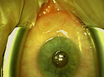
The patient, a 38-year-old woman, had recurrent eyelid swelling in both eyes and conjunctival inflammation with watery discharge beginning in June 2011 (Technical Appendix Figure, panel A). She visited numerous physicians and, upon various putative diagnoses (ranging from sicca syndrome to burnout syndrome), she received corresponding therapies, including antibiotics, steroids, and acupuncture. From January 2012 on, the eyelid swellings were accompanied by a creeping sensation and occurred more often. In early August 2012, she sought care at the emergency department of a university eye clinic in Vienna, Austria. She had a moving object in her left eye. Slit lamp examination revealed a white slender worm moving subconjunctivally in the temporal part of the left eye (Technical Appendix Figure, panel B). The conjunctiva was opened under topical anesthesia, and a 13-cm worm (Technical Appendix Figure, panel C) was removed (Video) and morphologically identified as a nongravid female of D. repens (Technical Appendix, panel D). Results of serologic testing for filariae were negative before and after extraction of the worm, as were results for testing of EDTA blood for microfilariae. Blood test results, including differential blood counts, were within reference ranges throughout the case history. The patient had returned from a 7-week stay in India, including the areas of Goa, Maharashtra, Delhi, and Uttar Pradesh, 3 months before initial onset of symptoms. Her travel history of the preceding 3 years included 4 more trips to India of several weeks each; a 2-week stay in Israel (October 2010); and a 2-week stay in Dubai, United Arab Emirates (July 2009).
For confirmation of the morphologic identification, we isolated DNA from a 1-cm piece of the worm after homogenization by using the QIAamp DNA Mini Kit (QIAGEN, Hilden, Germany). We amplified fragments of the cytochrome C oxidase subunit I (COI) with panfilarial primers COXfw 5′-GCKTTTCCTCGTGTTATGC-3′/COXrev 5′-CCAGCCAAAACAGGAACAG-3′ and 12S rRNA with panfilarial primers Panfil-12S-F 5′-gttccagaataatcggcta-3′/Panfil-12S-R 5′-attgacggatgrtttgtacc-3′ (7). We sequenced amplicons and subjected them to phylogenetic analyses (online Technical Appendix). All sequence data were submitted to GenBank (accession nos. KY750548–KY750550).
Figure

Figure. Phylogenetic analysis of the genus Dirofilaria based on cytochrome C oxidase subunit I gene sequences from a worm surgically extracted from the eye of a patient who had returned to Austria...
The 329 bp COI fragment (accession no. KY750548) showed 99%–100% identity to 2 sequences from Candidatus Dirofilaria hongkongensis (accession nos. KX265050 and JX187591). Identity to D. repens sequences was 95%–96%, to D. immitis 89%, and to Onchocerca spp. up to 92%. The 466 bp mitochondrial 12S rDNA fragment (accession no. KY750549) showed 99% identity to Candidatus Dirofilaria hongkongensis sequences from case-patients in India (accession no. KX265050) and Hong Kong (accession no. KY750550), the latter derived from original material of the first description of Candidatus Dirofilaria hongkongensis (6). Identity to a Dirofilaria sp. from a patient returning from India and Sri Lanka and to Dirofilaria sp. Thailand II, recently reported among dogs in Thailand (accession nos. KX265092 and KX265093) (8), was also 99%. Phylogenetic analysis using the COI sequence clearly placed the sequence into the Candidatus Dirofilaria hongkongensis cluster, the sister taxon to D. repens (Figure). Although D. immitis shows virtually identical COI sequences from 4 continents, genetic variability in D. repens–like parasites is obviously much higher, possibly associated with varying zoonotic potentials, reservoirs, and vectors; however, molecular data on Dirofilaria are still scarce.
In this case, Candidatus Dirofilaria hongkongensis was most likely acquired in India. An infection in Austria seems unlikely because, until now, only 1 singular autochthonous Dirofilaria infection has been reported, and that case was classic D. repens infection (4). Dubai is considered nonendemic for Dirofilaria spp. parasites, whereas Israel is known to be endemic for D. repens nematodes (1,3), but the patient’s trips to these countries were much longer ago than her latest trip to India. Moreover, all cases from India or Sri Lanka analyzed by us so far represented Candidatus Dirofilaria hongkongensis (8,9; S. Poppert, unpub. data), suggesting that this species is widely distributed on the Indian subcontinent. In fact, whether classical D. repens infection occurs in India at all is unclear. Infections with Candidatus Dirofilaria hongkongensis nematodes might take a similar course as infections with classical D. repens; however, a case of meningoencephalitis caused by nematodes of this candidate species also has been described (9). Dirofilaria spp. parasites isolated from human case-patients should be investigated by molecular methods to establish an exact species diagnosis, especially if infections were acquired outside Europe.
Dr. Winkler is a specialist for infectious diseases and tropical medicine at the General Hospital in Vienna, Austria. He is also professor of internal medicine at the Medical University of Vienna.
Acknowledgments
We thank Iveta Häfeli for excellent technical assistance and the Medical University of Vienna for financial support.
Informed written consent was obtained from the patient for publication of this study and any accompanying images and videos. We are very grateful to the patient for kindly providing all the material.
References
- Pampiglione S, Rivasi F. Human dirofilariasis due to Dirofilaria (Nochtiella) repens: an update of world literature from 1995 to 2000. Parassitologia. 2000;42:231–54.PubMedGoogle Scholar
- Pampiglione S, Rivasi F, Angeli G, Boldorini R, Incensati RM, Pastormerlo M, et al. Dirofilariasis due to Dirofilaria repens in Italy, an emergent zoonosis: report of 60 new cases. Histopathology. 2001;38:344–54. DOIPubMedGoogle Scholar
- Genchi C, Kramer LH, Rivasi F. Dirofilarial infections in Europe. Vector Borne Zoonotic Dis. 2011;11:1307–17. DOIPubMedGoogle Scholar
- Auer H, Susani M. The first autochthonous case of subcutaneous dirofilariosis in Austria [in German]. Wien Klin Wochenschr. 2008;120(19–20 Suppl 4):104‒6. https://doi.org/10.1007/s00508-008-1031-4PubMedGoogle Scholar
- Silbermayr K, Eigner B, Joachim A, Duscher GG, Seidel B, Allerberger F, et al. Autochthonous Dirofilaria repens in Austria. Parasit Vectors. 2014;7:226. DOIPubMedGoogle Scholar
- To KK, Wong SS, Poon RW, Trendell-Smith NJ, Ngan AH, Lam JW, et al. A novel Dirofilaria species causing human and canine infections in Hong Kong. J Clin Microbiol. 2012;50:3534–41. DOIPubMedGoogle Scholar
- Casiraghi M, Bain O, Guerrero R, Martin C, Pocacqua V, Gardner SL, et al. Mapping the presence of Wolbachia pipientis on the phylogeny of filarial nematodes: evidence for symbiont loss during evolution. Int J Parasitol. 2004;34:191–203. DOIPubMedGoogle Scholar
- Yilmaz E, Fritzenwanker M, Pantchev N, Lendner M, Wongkamchai S, Otranto D, et al. The mitochondrial genomes of the zoonotic canine filarial parasites Dirofilaria (Nochtiella) repens and Candidatus Dirofilaria (Nochtiella) hongkongensis provide evidence for presence of cryptic species. PLoS Negl Trop Dis. 2016;10:e0005028. DOIPubMedGoogle Scholar
- Poppert S, Hodapp M, Krueger A, Hegasy G, Niesen WD, Kern WV, et al. Dirofilaria repens infection and concomitant meningoencephalitis. Emerg Infect Dis. 2009;15:1844–6. DOIPubMedGoogle Scholar
Figures
Cite This ArticleTable of Contents – Volume 23, Number 8—August 2017
| EID Search Options |
|---|
|
|
|
|
|
|
Please use the form below to submit correspondence to the authors or contact them at the following address:
Julia Walochnik, Medical University of Vienna – Parasitology, Kinderspitalgasse 15, Vienna 1095, Austria
Top