Volume 24, Number 8—August 2018
Research
Susceptibility of Human Prion Protein to Conversion by Chronic Wasting Disease Prions
Figure 3
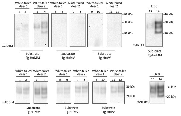
Figure 3. Evaluation of the in vitro conversion of human prion protein (PrP) seeded with the misfolded, disease-associated prion protein form present in chronic wasting disease (CWD)–affected white-tailed deer brain samples. We incubated 2 white-tailed deer CWD brain homogenates, derived from 2 affected animals (white-tailed deer 1 and 2), in a panel of 3 humanized transgenic substrates (Tg-HuMM, Tg-HuMV, and Tg-HuVV) and subjected them to a single round of protein misfolding cyclic amplification (PMCA) followed by proteinase K digestion. We diluted CWD brain homogenate 1:3 in PMCA substrate and performed Western blot analysis by using the mAb 3F4 (for the detection of human protease-resistant prion protein [PrPres]) and mAb 6H4 (for detection of CWD PrPres and human PrPres). We incorporated the elk specimen designated elk 0 as a control. We performed >3 repeats for the amplification white-tailed deer CWD 1 and 2 specimens with similar results. Reference molecular markers have been included. Molecular mass of electrophoretic markers is given. Odd and even number lanes show reaction mixtures before and after PMCA. mAb, monoclonal antibody; Tg-HuMM, humanized transgenic PRNP codon 129 homozygous methionine; Tg-Hu-MV, humanized transgenic methionine/valine; Tg-HuVV, humanized transgenic valine/valine.