Volume 25, Number 5—May 2019
Research
Lassa Virus Targeting of Anterior Uvea and Endothelium of Cornea and Conjunctiva in Eye of Guinea Pig Model
Figure 2

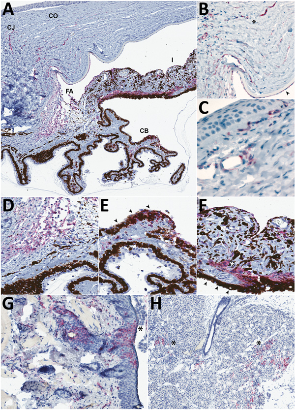
Figure 2. Detection of Lassa virus (LASV) antigen in the anterior uvea and endothelium within the eye of guinea pigs infected with LASV-Josiah and in the epithelium of structures adjacent to the eye in study of LASV targeting of anterior uvea and endothelium of cornea and conjunctiva in eye. A) Anterior uvea with LASV antigen immunolabeled (red) within the peripheral CO and CJ vessels, the FA, CB, and I. Original magnification ×4. B) Immunohistochemical (IHC) staining in the endothelium and adjacent stroma of the corneal margin (asterisk) and in the endothelium deep to Descemet’s membrane (arrowhead). Original magnification ×20 with 1.25 Optivar. C) Perivascular and endothelial staining in the bulbar conjunctiva. Original magnification ×63. D) IHC staining in the filtration angle. Original magnification ×20. E) Photomicrograph of the ciliary body highlighting the labeling in the pigmented epithelium (arrowheads) and stroma. Original magnification ×30. F) Photomicrograph of the iris showing IHC staining of LASV antigen in the stroma, smooth muscle (dilator muscle, white asterisk), and posterior pigmented epithelium (arrowheads). Original magnification ×40. G) IHC staining in eyelid epithelium (asterisk) and dermal vessels in the eyelid. Representative animal Jos-9. Original magnification ×15. H) IHC staining for LASV antigen in the acini of the lacrimal gland (asterisks). Representative animal Jos-9. Original magnification ×5. Representative animals: A–F, Jos-4; F, G, Jos-9. CB, ciliary body; CJ, conjunctival; CO, corneal; FA, filtration angle; I, iris; IHC, immunohistochemical.