Volume 25, Number 5—May 2019
Research
Lassa Virus Targeting of Anterior Uvea and Endothelium of Cornea and Conjunctiva in Eye of Guinea Pig Model
Figure 5
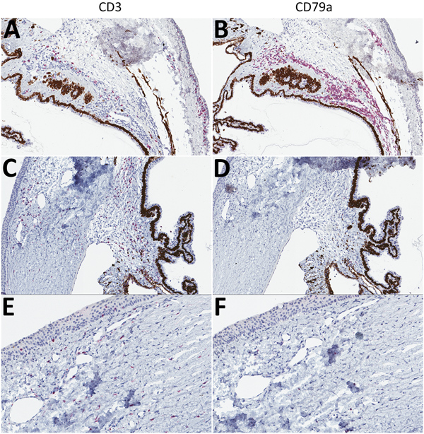
Figure 5. T-lymphocyte inflammation predominant in the eyes of animals that died of Lassa virus (LASV) infection >17 days postinfection in study of LASV targeting of anterior uvea and endothelium of cornea and conjunctiva in eye. CD3+ (left) and CD79a+ (right) lymphocyte antigens targeted by immunohistochemical (IHC) analysis are stained red. A) Inflamed filtration angle and sclera highlighting CD3+ T-lymphocytes. Original magnification ×10. B) Inflamed filtration angle and sclera highlighting the predominant population of CD79a+ B-lymphocytes. Original magnification ×10. C) Mildly inflamed filtration angle and sclera showing the predominance of CD3+ T-lymphocytes within the region. Original magnification ×10. D) Absence of CD79a+ B-lymphocytes. Original magnification ×10. E) New vessel formation at the margin of the cornea, indicating scattered CD3+ T-lymphocytes. Original magnification ×20. F) Minimal CD79a+ B-lymphocytes. Original magnification ×20. Representative animals: A, B, Jos-1; C–F, Jos-5.