Skip directly to site content Skip directly to page options Skip directly to A-Z link Skip directly to A-Z link Skip directly to A-Z link
Volume 25, Number 6—June 2019
Synopsis

Immunopathology of Fatal Human Variegated Squirrel Bornavirus 1 Encephalitis, Germany, 2011–2013

Dennis TappeComments to Author , Jonas Schmidt-Chanasit, Jessica Rauch, Petra Allartz, and Christiane Herden
Author affiliations: Bernhard Nocht Institute for Tropical Medicine, Hamburg, Germany (D. Tappe, J. Schmidt-Chanasit, J. Rauch, P. Allartz); German Centre for Infection Research, Hamburg (J. Schmidt-Chanasit); University of Giessen, Giessen, Germany (C. Herden)

Main Article

Figure 1

Brain inflammation in patients with fatal variegated squirrel bornavirus 1 encephalitis. A) Brain section showing mononuclear cell infiltration and tissue edema. Hematoxylin and eosin stain; original magnification ×100. B) Depiction of glial cell activation in a brain biopsy sample of malacia. Astrocytes appear bizarre and enlarged. Hematoxylin and eosin stain; original magnification ×400. C) Demonstration of glial fibrillary acidic protein (GFAP). Immunoperoxidase stain with hematoxylin counter

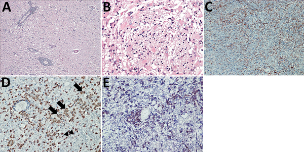
Figure 1. Brain inflammation in patients with fatal variegated squirrel bornavirus 1 encephalitis. A) Brain section showing mononuclear cell infiltration and tissue edema. Hematoxylin and eosin stain; original magnification ×100. B) Depiction of glial cell activation in a brain biopsy sample of malacia. Astrocytes appear bizarre and enlarged. Hematoxylin and eosin stain; original magnification ×400. C) Demonstration of glial fibrillary acidic protein (GFAP). Immunoperoxidase stain with hematoxylin counterstain; original magnification ×200. D) Predominantly foamy cells (lipid-laden microglia or macrophages [arrows]) and a few bushy microglia (arrowheads) as shown by marked CD68 positivity throughout the inflamed regions. Immunoperoxidase stain with hematoxylin counterstain; original magnification ×200. E) HLA-DR (brown) and CD68 positivity (blue) indicating many reactive microglia and macrophages. Immunoperoxidase and immunophosphatase stains; original magnification ×200.

Main Article

CrossRef reports the first page should be "A793", not "d541". (Ref. 8 "Stitz, Bilzer, Planz, 2002")

CrossRef reports the first page should be "1", not "379817". (Ref. 17 "Khan, Ahmad, Alshammari, Adnan, Baig, Lohani, et al., 2015")

Page created: May 20, 2019
Page updated: May 20, 2019
Page reviewed: May 20, 2019
The conclusions, findings, and opinions expressed by authors contributing to this journal do not necessarily reflect the official position of the U.S. Department of Health and Human Services, the Public Health Service, the Centers for Disease Control and Prevention, or the authors' affiliated institutions. Use of trade names is for identification only and does not imply endorsement by any of the groups named above.
file_external