Volume 29, Number 4—April 2023
Research Letter
Experimental Infection of North American Deer Mice with Clade I and II Monkeypox Virus Isolates
Cite This Article
Citation for Media
Abstract
The global spread of monkeypox virus has raised concerns over the establishment of novel enzootic reservoirs in expanded geographic regions. We demonstrate that although deer mice are permissive to experimental infection with clade I and II monkeypox viruses, the infection is short-lived and has limited capability for active transmission.
Monkeypox virus (MPXV; genus Orthopoxvirus, Poxviridae), which causes mpox disease, is a zoonotic pathogen that is endemic in Central Africa (clade I) and Western Africa (clade II) (1). In mid-May 2022, the World Health Organization first reported an increasing number of mpox cases in nonendemic countries, most of which had no established travel links to endemic regions (2). By October 2022, the outbreak encompassed >100 countries with reported confirmed mpox cases (3).
The global spread of MPXV outside of regions in which this virus was known to be endemic raises concerns over reverse zoonotic events resulting in the establishment of novel wildlife reservoirs. Small mammals, including rodents, have previously been implicated as enzootic reservoirs of MPXV. In North America, studies have shown that prairie dogs are susceptible to MPXV infection and may serve as a potential reservoir, but data on other wild rodents are limited (4). Peromyscus species rodents have an extensive and geographically diverse host range spanning most regions across North America and are well-established reservoirs for several zoonotic pathogens (5).
We evaluated the competency of deer mice (Peromyscus maniculatus rufinus) as a potential zoonotic reservoir for MPXV by using representative isolates from both clades. We infected groups of 12 adult (>6 weeks of age) deer mice with 1 of 3 MPXV isolates through intranasal instillation. The isolates included a clade II human isolate from the 2022 outbreak (MPXV/SP2833) (challenge dose 106 PFU); a second clade II virus isolated directly from a North American prairie dog (USA-2003) (challenge dose 106 PFU); and a historical clade I isolate (MPXV/V79-1-005) (challenge dose 104 PFU). For each virus preparation, we administered the maximum challenge dose based on titration on Vero cells. On days 4 and 10 postinfection, we euthanized 3 male and 3 female mice and collected selected solid organs for analysis of viral titers using molecular assays targeting of envelope protein gene (B6R) (6) and infectious viral quantification assays. In addition, we collected oral and rectal swab specimens and tested them similarly to assess the potential for shedding.
We conducted animal studies in accordance with the Canadian Council of Animal Care guidelines and following an animal use document approved by an institutional Animal Care and Use Committee, in a Biosafety Level 4 laboratory of the Public Health Agency of Canada. We conducted fully validated molecular assays in accordance with Public Health Agency of Canada special pathogens diagnostic procedures.
Figure
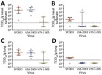
Figure. Monkeypox virus infectious titers from lung and nasal turbinate samples from experimentally infected deer mice. Groups of 12 deer mice (6 male, 6 female) were experimentally infected with monkeypox virus...
Throughout the course of the study, we observed no obvious signs of disease in any of the infected deer mice. We did not record daily weights because of the requirement for anesthetizing animals before any hands-on manipulation. Analysis of tissue samples from mice infected with the 2022 Canada isolate (MPXV/SP2833) revealed limited and sporadic spread of MPXV beyond the sites of inoculation (nasal turbinates and lungs) (Table). By comparison, USA-2003 appeared to disseminate beyond the respiratory tract, resulting in uniform detection of MPXV DNA in liver and spleen specimens collected at 4 days postinfection (dpi). The clade I virus (MPXV/V79-1-005) yielded results more similar to those for USA-2003; nasal turbinate, lung, liver and spleen samples were positive at 4 dpi. By day 10 dpi, organ specimens from most mice across the 3 infection groups were trending toward clearance (Table). Infectious titers conducted on lung and nasal turbinate specimens collected at both timepoints from the 3 challenge groups corroborated these findings and demonstrated decreasing viral titers between the 2 timepoints (Figure).
Of note, the clade I virus did not achieve high titers in either organ, even when analyzed at 4 dpi. Although this finding may suggest the MPXV/V79-1-005 isolate does not replicate as efficiently in deer mice, the apparent low viral titers observed may be attributable to the lower inoculum dose. A similar challenge dose of this strain resulted in lethal infection in CAST/EiJ mice (7). Further, subsequent cell culture propagations of MPXV/V79-1-005 resulted in similar titers as the clade II isolates used previously, suggesting that all 3 replicate to a similar extent on Vero cells. Nevertheless, follow-up studies with other clade I viruses are warranted.
We collected oral and rectal swab specimens to assess shedding and the potential for transmission of MPXV from infected deer mice. Overall, shedding, as suggested by the presence of MPXV DNA in swab extracts, was readily detectable in deer mice inoculated with either clade II virus at day 4, but we noted decreasing levels of positivity by day 10. Shedding of MPXV/V79-1-005 (clade 1) was far less than that of either of the clade II viruses we evaluated (Table).
Our study suggests that these rodents may support a short-term but abortive infection with at least clade II MPXV isolates, although with limited capacity to spread. Given the short duration of infection, these animals probably do not represent a viable enzootic reservoir for MPXV. Further studies should be conducted on other rodents in North America and Europe to assess their competency as vectors or reservoirs of MPXV. Particular interest should be given to Rattus species rodents that may frequently come into contact with medical waste containing viable MPXV.
Mr. Deschambault is a senior laboratory technician in the Special Pathogens Program of the Public Health Agency of Canada. His research interests include disease modeling and vaccine development for emerging and high-consequence viral pathogens.
Acknowledgments
This work was funded by the Public Health Agency of Canada. We obtained the MPXV USA-2003 reagent NR-2500 through the BEI Resources Repository, National Institute of Allergy and Infectious Diseases, National Institutes of Health.
All authors declare no conflict of interest.
References
- Likos AM, Sammons SA, Olson VA, Frace AM, Li Y, Olsen-Rasmussen M, et al. A tale of two clades: monkeypox viruses. J Gen Virol. 2005;86:2661–72. DOIPubMedGoogle Scholar
- Velavan TP, Meyer CG. Monkeypox 2022 outbreak: An update. Trop Med Int Health. 2022;27:604–5. DOIPubMedGoogle Scholar
- Centers for Disease Control and Prevention. 2022 monkeypox outbreak global map [cited 2022 Oct 18]. https://www.cdc.gov/poxvirus/monkeypox/response/2022/world-map.html
- Parker S, Buller RM. A review of experimental and natural infections of animals with monkeypox virus between 1958 and 2012. Future Virol. 2013;8:129–57. DOIPubMedGoogle Scholar
- Barbour AG. Infection resistance and tolerance in Peromyscus spp., natural reservoirs of microbes that are virulent for humans. Semin Cell Dev Biol. 2017;61:115–22. DOIPubMedGoogle Scholar
- Li Y, Olson VA, Laue T, Laker MT, Damon IK. Detection of monkeypox virus with real-time PCR assays. J Clin Virol. 2006;36:194–203. DOIPubMedGoogle Scholar
- Warner BM, Klassen L, Sloan A, Deschambault Y, Soule G, Banadyga L, et al. In vitro and in vivo efficacy of tecovirimat against a recently emerged 2022 monkeypox virus isolate. Sci Transl Med. 2022;14:
eade7646 . DOIPubMedGoogle Scholar
Figure
Table
Cite This ArticleOriginal Publication Date: March 06, 2023
Table of Contents – Volume 29, Number 4—April 2023
| EID Search Options |
|---|
|
|
|
|
|
|
Please use the form below to submit correspondence to the authors or contact them at the following address:
David Safronetz, Special Pathogens Program, National Microbiology Laboratory Branch, Public Health Agency of Canada, 1015 Arlington St, Winnipeg, MB R3E 3R2, Canada
Top