Skip directly to site content Skip directly to page options Skip directly to A-Z link Skip directly to A-Z link Skip directly to A-Z link

Disclaimer: Early release articles are not considered as final versions. Any changes will be reflected in the online version in the month the article is officially released.

Volume 30, Number 7—July 2024
Research

Highly Pathogenic Avian Influenza A(H5N1) Clade 2.3.4.4b Virus Infection in Domestic Dairy Cattle and Cats, United States, 2024

Eric R. BurroughComments to Author , Drew R. Magstadt, Barbara Petersen, Simon J. Timmermans, Phillip C. Gauger, Jianqiang Zhang, Chris Siepker, Marta Mainenti, Ganwu Li, Alexis C. Thompson, Patrick J. Gorden, Paul J. Plummer, and Rodger Main
Author affiliations: Iowa State University College of Veterinary Medicine, Ames, Iowa, USA (E.R. Burrough, D.R. Magstadt, P.C. Gauger, J. Zhang, C. Siepker, M. Mainenti, G. Li, P.J. Gorden, P.J. Plummer, R. Main); Sunrise Veterinary Service PLLC, Amarillo, Texas, USA (B. Petersen); Veterinary Research & Consulting Services LLC, Hays, Kansas, USA (S.J. Timmermans); Texas A&M Veterinary Medical Diagnostic Laboratory, College Station, Texas, USA (A.C. Thompson)

Main Article

Figure 1

Mammary gland lesions in cattle in study of highly pathogenic avian influenza A(H5N1) clade 2.3.4.4b virus infection in domestic dairy cattle and cats, United States, 2024. A, B) Mammary gland tissue sections stained with hematoxylin and eosin. A) Arrowheads indicate segmental loss within open secretory mammary alveoli. Original magnification ×40. B) Arrowheads indicate epithelial degeneration and necrosis lining alveoli with intraluminal sloughing. Asterisk indicates intraluminal neutrophilic inflammation. Original magnification ×400. C, D) Mammary gland tissue sections stained by using avian influenza A immunohistochemistry. C) Brown staining indicates lobular distribution of avian influenza A virus. Original magnification ×40. D) Brown staining indicates strong nuclear and intracytoplasmic immunoreactivity of intact and sloughed epithelial cells within mammary alveoli. Original magnification ×400.

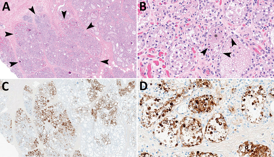
Figure 1. Mammary gland lesions in cattle in study of highly pathogenic avian influenza A(H5N1) clade 2.3.4.4b virus infection in domestic dairy cattle and cats, United States, 2024. A, B) Mammary gland tissue sections stained with hematoxylin and eosin. A) Arrowheads indicate segmental loss within open secretory mammary alveoli. Original magnification ×40. B) Arrowheads indicate epithelial degeneration and necrosis lining alveoli with intraluminal sloughing. Asterisk indicates intraluminal neutrophilic inflammation. Original magnification ×400. C, D) Mammary gland tissue sections stained by using avian influenza A immunohistochemistry. C) Brown staining indicates lobular distribution of avian influenza A virus. Original magnification ×40. D) Brown staining indicates strong nuclear and intracytoplasmic immunoreactivity of intact and sloughed epithelial cells within mammary alveoli. Original magnification ×400.

Main Article

Page created: April 29, 2024
Page updated: April 29, 2024
Page reviewed: April 29, 2024
The conclusions, findings, and opinions expressed by authors contributing to this journal do not necessarily reflect the official position of the U.S. Department of Health and Human Services, the Public Health Service, the Centers for Disease Control and Prevention, or the authors' affiliated institutions. Use of trade names is for identification only and does not imply endorsement by any of the groups named above.
file_external