Volume 30, Number 7—July 2024
Research
Highly Pathogenic Avian Influenza A(H5N1) Clade 2.3.4.4b Virus Infection in Domestic Dairy Cattle and Cats, United States, 2024
Figure 2
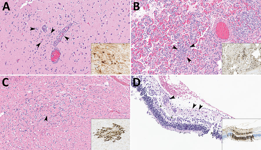
Figure 2. Lesions in cat tissues in study of highly pathogenic avian influenza A(H5N1) clade 2.3.4.4b virus infection in domestic dairy cattle and cats, United States, 2024. Tissue sections were stained with hematoxylin and eosin; insets show brown staining of avian influenza A viruses via immunohistochemistry by using the chromogen 3,3′-diaminobenzidine tetrahydrochloride. Original magnification ×200 for all images and insets. A) Section from cerebral tissue. Arrowheads show perivascular lymphocytic encephalitis, gliosis, and neuronal necrosis. Inset shows neurons. B) Section of lung tissue showing lymphocytic and fibrinous interstitial pneumonia with septal necrosis and alveolar edema; arrowheads indicate lymphocytes. Inset shows bronchiolar epithelium, necrotic cells, and intraseptal mononuclear cells. C) Section of heart tissue. Arrowhead shows interstitial lymphocytic myocarditis and focal peracute myocardial coagulative necrosis. Inset shows cardiomyocytes. D) Section of retinal tissue. Arrowheads show perivascular lymphocytic retinitis with segmental neuronal loss and rarefaction in the ganglion cell layer. Asterisks indicate attenuation of the inner plexiform and nuclear layers with artifactual retinal detachment. Insets shows all layers of the retina segmentally within affected areas have strong cytoplasmic and nuclear immunoreactivity to influenza A virus.