SARS-CoV-2 Seropositivity in Urban Population of Wild Fallow Deer, Dublin, Ireland, 2020–2022
Kevin Purves
1, Hannah Brown
1, Ruth Haverty, Andrew Ryan, Laura L. Griffin, Janet McCormack, Sophie O’Reilly, Patrick W. Mallon, Virginie Gautier, Joseph P. Cassidy, Aurelie Fabre, Michael J. Carr, Gabriel Gonzalez, Simone Ciuti, and Nicola F. Fletcher

Author affiliations: University College Dublin, Dublin, Ireland (K. Purves, H. Brown, R. Haverty, A. Ryan, L.L. Griffin, J. McCormack, S. O’Reilly, P.W. Mallon, V. Gautier, J.P. Cassidy, A. Fabre, M.J. Carr, G. Gonzalez, S. Ciuti, N.F. Fletcher); St Vincent's University Hospital, Dublin (P.W. Mallon, A. Fabre); Hokkaido University, Sapporo, Japan (M.J. Carr, G. Gonzalez)
Main Article
Figure 11
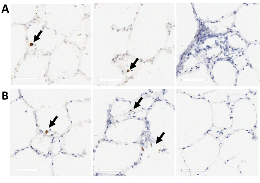
Figure 11. SARS-CoV-2 Omicron BA.1 infection of ex vivo lung tissue in study of SARS-CoV-2 seropositivity in urban population of wild fallow deer, Dublin, Ireland, 2020–2022. Precision cut lung slices were collected from 2 SARS-CoV-2–seronegative deer and inoculated with SARS-CoV-2 Omicron BA.1; sections were stained by using immunohistochemistry. Control sections were stained with IgG only or mock infected. A) Deer 1; B) deer 2. Arrows in first and middle panels indicate Omicron BA.1 immunoreactivity in cells morphologically consistent with type 2 pneumocytes. Third panel indicates no immunoreactivity after staining with the IgG control. No immunoreactivity was observed in the mock-infected tissues for either animal. Scale bars indicate 60 μm.
Main Article
Page created: June 03, 2024
Page updated: July 20, 2024
Page reviewed: July 20, 2024
The conclusions, findings, and opinions expressed by authors contributing to this journal do not necessarily reflect the official position of the U.S. Department of Health and Human Services, the Public Health Service, the Centers for Disease Control and Prevention, or the authors' affiliated institutions. Use of trade names is for identification only and does not imply endorsement by any of the groups named above.