Skip directly to site content Skip directly to page options Skip directly to A-Z link Skip directly to A-Z link Skip directly to A-Z link

Disclaimer: Early release articles are not considered as final versions. Any changes will be reflected in the online version in the month the article is officially released.

Volume 30, Number 9—September 2024
Dispatch

Avian and Human Influenza A Virus Receptors in Bovine Mammary Gland

Charlotte KristensenComments to Author , Henrik E. Jensen, Ramona Trebbien, Richard J. Webby, and Lars E. Larsen
Author affiliations: University of Copenhagen, Frederiksberg, Denmark (C. Kristensen, H.E. Jensen, L.E. Larsen); Statens Serum Institut, Copenhagen, Denmark (R. Trebbien); St. Jude Children’s Research Hospital, Memphis, Tennessee, USA (R.J. Webby)

Main Article

Figure 1

Results of staining showing wide expression of the human receptor for influenza A virus (designated SNA) in the bovine mammary gland. A) An example of the SNA staining of a mammary gland from a cow in late lactation. Arrowhead indicates expression of the human receptor in the active alveoli. Asterisks indicate less staining in the less active alveoli. Original magnification ×10. B, C) SNA staining of a duct (B) and (C) an alveolus in a 7-year-old cow. Original magnification ×60. D, E) Positive staining obtained from the image analysis. Green line shows the region of interest. Original magnification ×60. F) A neuraminidase pretreatment negative control showed markedly reduced staining of the SNA lectin. Original magnification ×60. The staining was visualized using Vector Blue (Vector Laboratories, https://vectorlabs.com).

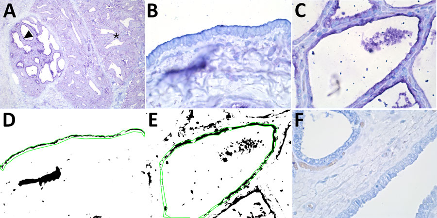
Figure 1. Results of staining showing wide expression of the human receptor for influenza A virus (designated SNA) in the bovine mammary gland. A) An example of the SNA staining of a mammary gland from a cow in late lactation. Arrowhead indicates expression of the human receptor in the active alveoli. Asterisks indicate less staining in the less active alveoli. Original magnification ×10. B, C) SNA staining of a duct (B) and (C) an alveolus in a 7-year-old cow. Original magnification ×60. D, E) Positive staining obtained from the image analysis. Green line shows the region of interest. Original magnification ×60. F) A neuraminidase pretreatment negative control showed markedly reduced staining of the SNA lectin. Original magnification ×60. The staining was visualized using Vector Blue (Vector Laboratories, https://vectorlabs.com).

Main Article

Page created: July 17, 2024
Page updated: August 09, 2024
Page reviewed: August 09, 2024
The conclusions, findings, and opinions expressed by authors contributing to this journal do not necessarily reflect the official position of the U.S. Department of Health and Human Services, the Public Health Service, the Centers for Disease Control and Prevention, or the authors' affiliated institutions. Use of trade names is for identification only and does not imply endorsement by any of the groups named above.
file_external