Volume 31, Number 11—November 2025
Research
Isolation and Characterization of Rickettsia finnyi, Novel Pathogenic Spotted Fever Group Rickettsia in Dogs, United States
Praveen K. Korla1, Michael G. Karounos1, Sarah B. Clarke, Cynthia Robveille, James M. Wilson, Edward B. Breitschwerdt, Adam J. Birkenheuer, and Barbara A. Qurollo
Figure 3
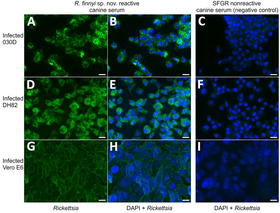
Figure 3. Microscopic images from study of isolation and characterization of Rickettsia finnyi, novel pathogenic spotted fever group Rickettsia in dogs, United States. Images depict R. finnyi sp. nov. strain 2024-CO-Wats–infected 030D canine mononuclear cells (A–C), DH82 canine histiocytic cells (D–F), and Vero E6 primate epithelial cells (G–I) detected by immunofluorescence using R. finnyi sp. nov.–seroreactive canine serum (dog 2, May 15 date from Table 2). Scale bar indicates 20 µm. SFGR nonreactive canine serum was used as a negative control. Green represents 2024-CO-Wats organisms. Blue represents nuclei of individual mammalian host cells (DAPI). SFGR, spotted fever group Rickettsia.
1These authors contributed equally to this article.
Page created: September 24, 2025
Page updated: December 10, 2025
Page reviewed: December 10, 2025
The conclusions, findings, and opinions expressed by authors contributing to this journal do not necessarily reflect the official position of the U.S. Department of Health and Human Services, the Public Health Service, the Centers for Disease Control and Prevention, or the authors' affiliated institutions. Use of trade names is for identification only and does not imply endorsement by any of the groups named above.