Volume 31, Supplement—May 2025
SUPPLEMENT ISSUE
Supplement
16S Ribosomal RNA Gene PCR and Sequencing for Pediatric Infection Diagnosis, United States, 2020–2023
Cite This Article
Citation for Media
Abstract
Gene PCR and sequencing using 16S ribosomal RNA (rRNA) can help diagnose challenging bacterial infections. Data on the optimal clinical settings for this type of testing are limited. We performed a retrospective study at Mayo Clinic, Rochester, Minnesota, USA, with typically sterile specimens from children that underwent 16S rRNA PCR testing during September 2020–December 2023. Of 162 tests performed on 124 patients, 20% were positive; 58% of positive samples were from culture-negative specimens. Fluid specimens were >3 times as likely to test positive as tissue specimens (odds ratio 3.07 [95% CI 1.32–7.11]; p = 0.007), and pleural fluid demonstrated the highest positivity rate (50%). Of 33 positive results, 4 (12%) specimens qualified for reporting to the state health department for communicable diseases. Those single-laboratory findings demonstrate that the highest positivity rate of 16S rRNA PCR and sequencing is pleural fluid, although many specimen types tested positive.
Gene PCR using 16S ribosomal RNA (rRNA) followed by sequencing can identify bacteria in normally sterile body tissues and fluids (1,2). This method may serve as a diagnostic tool in complex bacterial infections, particularly when conventional tests fail to identify pathogens (3,4). The clinical use of 16S rRNA PCR and sequencing has been shown to yield concordant results with bacterial cultures (when positive), to enhance detection of fastidious bacteria, and to assist in antimicrobial drug stewardship (4–8). However, the diagnostic yield of 16S rRNA PCR and sequencing from various specimen sources has been variable in published studies (4,6,9–11); diagnostic yield may vary on the basis of patient and specimen characteristics. Data on optimal clinical settings and specimen selection for this testing are limited, particularly in pediatrics (9,12).
Mayo Clinic (Rochester, MN, USA) began offering 16S rRNA PCR and sequencing clinically in 2017; the sequencing initially involving Sanger sequencing alone (4). Then, in 2019, to increase positivity rates and to decatenate mixed sequences because of 16S rRNA gene copy variants or polymicrobial infections, next-generation sequencing (NGS) was substituted for or added to Sanger sequencing of the PCR-amplified 16S rRNA gene when needed (13). This study reviews Mayo Clinic’s clinical experience with 16S rRNA PCR and sequencing of specimens from children to identify clinical syndromes where this testing is useful and to optimize specimen choice.
Study Design
We performed a retrospective study involving specimens collected from Mayo Clinic patients 0–18 years of age whose normally sterile tissue or fluid specimens underwent 16S rRNA PCR and sequencing during September 2020–December 2023. We identified patients and 16S rRNA PCR and sequencing results by using the clinical microbiology laboratory database and collected demographic, clinical, and microbiologic data from the electronic medical record. If a patient had specimens collected from the same source during different encounters, we included only specimens from the first encounter. In routine clinical practice, holding a specimen in the clinical microbiology laboratory for 14 days for potential 16S rRNA PCR and sequencing, if clinically needed, was offered as an option. This study was approved by the Mayo Clinic Institutional Review Board (protocol no. 20–012373).
Definitions
Immunocompromised hosts included patients with malignancies undergoing chemotherapy, those who had undergone solid organ or hematopoietic stem cell transplantation, and those receiving high-dose steroids (pulse dose steroids 20 mg/d for >14 days, or dexamethasone for >10 days) or other immunosuppressive agents. We defined intensive care unit (ICU) admission as receiving medical care in the neonatal, pediatric, or cardiovascular ICU at the time of specimen collection.
We categorized cerebrospinal fluid, ovarian fluid, pericardial fluid, peritoneal fluid, pleural fluid, subdural fluid, synovial fluid, and vitreous fluid as fluid specimens and other specimens (e.g., bone) as tissue specimens. We collected the results of conventional testing, which included Gram stain, bacterial culture, BioFire Meningitis and Encephalitis (ME) panel (bioMérieux, https://www.biomerieux.com), and Kingella kingae PCR if clinically performed on specimens collected at the same time and from the same site as specimens for 16S rRNA PCR and sequencing. We calculated the turnaround time as the interval from specimen collection to result finalization. We defined prior antibacterial therapy as any antimicrobial drugs administered within 24 hours before the test order for 16S rRNA PCR and sequencing.
Specimen Processing
We performed specimen processing and bacterial culture in the Clinical Bacteriology Laboratories of the Division of Clinical Microbiology at Mayo Clinic. We identified isolated bacteria by using conventional biochemical methods or matrix-assisted laser desorption/ionization time-of-flight mass spectrometry. Details of the 16S rRNA PCR and sequencing procedure have been described previously (13). In brief, the assay involved an up-front real-time PCR assay, reported as negative or submitted to Sanger or NGS on the basis of cycle threshold (Ct) value. Specimens with Ct values <32 cycles underwent bidirectional Sanger sequencing by using an Applied Biosystems 3500xL Genetic Analyzer (Thermo Fisher Scientific, https://www.thermofisher.com). We sent specimens with Ct values of 32–34 or <32 with Sanger sequencing that yielded an uninterpretable result to NGS by using an Illumina MiSeq System (Illumina, https://www.illumina.com) with a 500-cycle (2 × 250 paired-end read) v2 nano kit. We reported specimens with Ct values >34 as negative, except if we observed a well-defined melting temperature peak (>0.4), in which case we sent them to NGS. We used Pathogenomix (https://www.pathogenomix.com) for quality control processes and the Pathogenomix PRIME database for sequence analysis. The Pathogenomix Prime database contains 48,139 curated 16S rRNA gene sequences. The processor filters low-quality reads (Q<30) and clusters sequences on the basis of >210-bp length, >100 copies, and 0% variation.
Statistical Analysis
We compared characteristics between positive and negative tests by using a 2-sample t-test for continuous variables. For categorical variables with >5 observations, we calculated odds ratios (ORs) and 95% CIs by using unconditional maximum likelihood estimation; we obtained p values by using χ2 tests. For categorical variables with <5 observations, we calculated ORs and 95% CIs by using conditional maximum likelihood estimation and obtained p values were by using Fisher exact tests. We considered a 2-tailed p value <0.05 statistically significant.
Patients
Figure
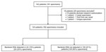
Figure. Specimen flowchart from a study of 16S ribosomal RNA gene PCR and sequencing for pediatric infection diagnosis, Mayo Clinic, Rochester, Minnesota, USA, 2020–2023. Specimens from tongue, ear canal, and nose...
A total of 124 pediatric patients with 162 tests from typically sterile sources were included (Figure). At sampling, 20% (n = 24) of patients were identified as immunocompromised hosts, and 37% (n = 46) of patients were in ICUs (Table 1). The most common suspected clinical manifestations were meningoencephalitis, musculoskeletal infection, and pleural effusion.
16S rRNA PCR and Sequencing Results
The mean turnaround time for positive 16S rRNA PCR and sequencing tests was 8 days (3.2–12.8 days), whereas for negative tests it was 3 days (0–6.7 days) (Table 2). A total of 84 (50%) specimens were collected from patients who received antimicrobial drugs within 24 hours before sampling, which was associated with a higher likelihood of positive results (p = 0.001).
The overall 16S rRNA PCR and sequencing positivity rate was 20% among all 162 specimens collected from 124 patients (Figure). Fluid specimens were 3-fold more likely to test positive compared with tissue specimens (OR 3.07 [95% CI 1.32–7.11]; p = 0.007) (Table 2). The most frequent specimen sources were cerebrospinal fluid, bone tissue, deep soft tissue, synovial fluid, and pleural fluid. Among those, specimens with high positivity rates included pleural fluid (50%, n = 5) and synovial fluid (43%, n = 9); there were no positive results from deep soft tissue specimens.
Among the 33 positive tests, 12 (36%) tests were polymicrobial detections. The most common single bacteria identified was Staphylococcus aureus complex in 4 (12%) positive tests, followed by Kingella kingae in 3 (9%) positive tests (all synovial fluid); other bacteria each accounted for 3%–9% of positive tests from various sources (Table 3). We recorded details of test results and clinical diagnoses for 24 patients with positive 16S rRNA PCR and sequencing results (Appendix Table).
Comparison to Conventional Tests
Among 152 specimens tested with both Gram stain and 16S rRNA PCR and sequencing, 21% (n = 7) of positive specimens had a corresponding positive Gram stain, whereas none of the negative tests were associated with a positive Gram stain. Patients with positive Gram stains had a higher likelihood of positive 16S rRNA PCR and sequencing results compared with patients with negative Gram stains (p<0.0001) (Table 4).
Of the 161 specimens tested with both bacterial cultures and 16S rRNA PCR and sequencing, 133 (83%) specimens demonstrated concordant results between the 2 methods: 14 (9%) specimens were positive after both tests and 119 (74%) specimens were negative after both tests. In addition, 19 specimens with negative bacterial cultures were positive by 16S rRNA PCR and sequencing: polymicrobial infections (n = 9), K. kingae (n = 3), Fusobacterium naviforme/nucleatum (n = 2), Streptococcus mitis group (n = 2), Cardiobacterium hominis (n = 1), Pseudomonas aeruginosa (n = 1), and Streptococcus pyogenes (n = 1).
Nine 16S rRNA PCR and sequencing tests were negative despite positive cultures: 4 positive results for Cutibacterium acnes from bacterial cultures in periimplant and bone tissue, 1 positive result for Staphylococcus capitis from bacterial culture in a bone tissue specimen, and 5 cases of suspected culture contamination. The contamination cases involved isolations of Staphylococcus epidermidis from pleural fluid (n = 1), deep soft tissue (n = 1), and lymph node tissue (n = 1) and Niallia circulans group from bone (n = 1) and deep soft tissue (n = 1).
Of the 23 specimens tested with both the BioFire ME panel and 16S rRNA PCR and sequencing, 2 were negative by the panel with positive 16S rRNA PCR and sequencing results (S. epidermidis and F. naviforme/nucleatum). The S. epidermidis case was considered a contaminant. No bacterial pathogens were identified by the BioFire ME panel that were not also detected by 16S rRNA PCR and sequencing.
Of the 7 synovial fluid specimens tested with both K. kingae PCR and 16S rRNA PCR and sequencing, 86% (n = 6) of specimens showed concordant positive or negative results. 16S rRNA PCR and sequencing detected K. kingae in 1 synovial fluid specimen that tested negative with synovial fluid K. kingae PCR.
Multiple Tests on the Same Specimen Type
At least 2 16S rRNA PCR and sequencing tests were ordered for 22 patients on the same specimen source during the same procedure (Table 5), mostly bone tissue, deep soft tissue, and synovial fluid. All tests yielded concordant results, either negative or positive.
Specimen Hold Strategy
Clinicians placed a request to hold a specimen for potential future 16S rRNA PCR and sequencing testing on 17 specimens (Table 2). Over the ensuing clinical course, because of positive Gram stains and negative bacterial cultures after 24–48 hours of incubation, 16S rRNA PCR and sequencing tests were performed on the saved specimens. Of those, 3 tests had positive 16S rRNA PCR and sequencing results, including identification of S. mitis group in 2 pleural fluid specimens and S. dysgalactiae in 1 synovial fluid specimen.
In this study, we conducted a 3-year retrospective evaluation of the diagnostic yield of 16S rRNA PCR and sequencing in children by using various specimen types. We were unable to find many other published studies exploring the application of 16S rRNA PCR and sequencing in pediatric patients. The overall test positivity rate we found was 20%, consistent with previous studies in pediatric patients, which reported positivity rates ranging from 14% to 23% (6,9,10,14). Initiation of empiric therapy within 24 hours before sampling did not negatively affect positivity rates, consistent with findings from studies in adults and children (4,9,11).
Subgroup analysis revealed that 16S rRNA PCR and sequencing had a higher positivity rate in fluid compared with tissue specimens, especially in pleural fluid, which provided additional diagnostic value for pathogens such as S. mitis group and S. pyogenes. Despite the limited pediatric sample size, our findings are consistent with prior studies indicating that pleural fluid yields a high positivity rate (10,14,15).
A potential limitation of our study is that bronchoalveolar lavage (BAL) fluid was not tested; in prior studies, BAL fluid has been reported as a common specimen source for 16S rRNA PCR and sequencing testing. However, despite high positivity rates in BAL fluids, the clinical relevance of those findings has been questionable (6,9,16), possibly because BAL fluid is not sterile. In contrast, sample dilution during bronchoscopy can increase the likelihood of false-negative results.
The yield of 16S rRNA PCR and sequencing in bone and joint infection has varied in previous research, ranging from 21% to 32% (17–19). Bone tissue and synovial fluids or tissues were the most common sources in this study. Compared with the single K. kingae PCR test on synovial fluid used at the Mayo Clinic, the 16S rRNA PCR and sequencing offered additional diagnostic value in only 1 of 7 cases. Given the shorter turnaround time of the K. kingae PCR test, a single PCR test remains the optimal first-line test for suspected bone and joint infections in toddlers. This target is also available on the BioFire joint infection (JI) panel (20). The BioFire JI panel has been used for rapid diagnosis of pediatric septic arthritis, offering a fast turnaround, and sensitive and specific detection of on-panel microorganisms and select antimicrobial resistance genes (20,21). Compared with the BioFire JI panel, 16S rRNA PCR and sequencing demonstrated higher sensitivity in periprosthetic JI (PJI) because the BioFire JI panel does not include S. epidermidis, a common cause of PJI (22–24).
We found discrepancies in C. acnes testing, in which cultures were positive but 16S rRNA PCR and sequencing was negative (4 peri-implant and bone tissue specimens). Those discrepancies are likely because of the limited ability to report low abundance C. acnes from 16S rRNA PCR and sequencing because of its frequent presence in background sequences, as published previously (25,26).
Of the 23 BioFire ME panels performed, 19 had concordant negative results by 16S rRNA PCR and sequencing, in keeping with other studies’ findings (4,27). 16S rRNA PCR and sequencing uniquely identified F. naviforme/nucleatum, which is not included in the ME panel (28). Two cases were negative by 16S rRNA PCR and sequencing but positive for viruses by the BioFire ME panel; this is expected because 16S rRNA PCR and sequencing targets bacterial DNA, while the BioFire ME panel includes viral targets. Turnaround time is a key factor to consider. Our findings underscore the value of using the BioFire ME panel ahead of 16S rRNA PCR and sequencing, proceeding to 16S rRNA PCR and sequencing when the BioFire ME panel is negative (29).
In this study, multiple 16S rRNA PCR and sequencing tests performed on specimens from the same specimen source collected during the same procedure resulted in no discordant results. Assessment of the clinical value of performing multiple 16S rRNA PCR and sequencing tests has been limited. A multicenter study on adult PJI showed that collecting 5 perioperative samples per patient for culture and 16S rRNA PCR and sequencing showed a lack of sensitivty of the latter in the diagnosis of PJI (30). Another report indicated that testing multiple samples per patient may help rule out potential contaminating microorganisms (31). Our findings indicate a single 16S rRNA PCR and sequencing test on 1 specimen, collected along with at >2 specimens for bacterial culture during the same procedure, may be adequate.
This study also explored the role of collecting and holding a specimen for future testing if clinically indicated. Positive detections were found in 3 cases managed with this strategy. We conceive that use of this diagnostic pathway could optimize testing resource use. Further research with larger sample sizes is necessary to determine the clinical syndromes and specimen sources that would benefit from delayed or reflexive testing.
The use of 16S rRNA PCR and sequencing in clinical practice has implications for public health, including enhanced detection of bacteria that may be notifiable infectious diseases. Clinical laboratories should establish protocols for reporting detected pathogens to public health authorities, and public health laboratories should define which molecularly detected species are reportable from which specimen types. As demonstrated in this study, K. kingae, often missed by conventional cultures, is readily detected by 16S rRNA PCR and sequencing. Clinical use of this assay can provide data useful for identifying outbreaks and informing timely public health interventions (32).
The first limitation of this study is that the small sample size limits statistical power. Second, the study was conducted at a single institution, limiting generalizability. Finally, subgroup analysis of suspected clinical syndromes and outcomes was not performed. Future studies with larger sample sizes, specimens collected from multiple sites, comprehensive clinical outcomes recorded, and adjustments for potential confounders are warranted.
In conclusion, this study demonstrates that 16S rRNA PCR and sequencing yields the highest positivity rate in fluid specimens, particularly pleural and synovial fluids from children. A strategy of collecting specimens for future testing, if clinically indicated, is described as a diagnostic stewardship tool. Further research should focus on optimizing use of the described testing use in conjunction with other testing, while considering overall turnaround time. Implementation research is needed to evaluate the effect of 16S rRNA PCR and sequencing on patient outcomes.
Dr. Li is a physician and assistant professor of pediatrics in the Division of Pediatric Infectious Diseases, Department of Pediatrics, at the Mayo Clinic. His primary research interests include novel molecular microbiological diagnostics in children and using phage therapy to treat multidrug-resistant bacteria.
Acknowledgment
Author contributions: G.L., C.A.R., and J.T.G. designed the study; G.L., C.A.R., R.M.K., and N.T.S. collected the data; G.L., Z.Z., and J.M. analyzed the data; M.J.W. and R.P. developed the assay; G.L., Z.Z., and R.M.K. wrote the manuscript; C.A.R., R.M.K., N.T.S., E.H.R., J.M., J.T.G., and R.P. revised the manuscript.
References
- Drevinek P, Hollweck R, Lorenz MG, Lustig M, Bjarnsholt T. Direct 16S/18S rRNA PCR followed by Sanger sequencing as a clinical diagnostic tool for detection of bacterial and fungal infections: a systematic review and meta-analysis. J Clin Microbiol. 2023;61:
e0033823 . DOIPubMedGoogle Scholar - Church DL, Cerutti L, Gürtler A, Griener T, Zelazny A, Emler S. Performance and application of 16S rRNA gene cycle sequencing for routine identification of bacteria in the clinical microbiology laboratory. Clin Microbiol Rev. 2020;33:e00053–19. DOIPubMedGoogle Scholar
- Rampini SK, Bloemberg GV, Keller PM, Büchler AC, Dollenmaier G, Speck RF, et al. Broad-range 16S rRNA gene polymerase chain reaction for diagnosis of culture-negative bacterial infections. Clin Infect Dis. 2011;53:1245–51. DOIPubMedGoogle Scholar
- Fida M, Khalil S, Abu Saleh O, Challener DW, Sohail MR, Yang JN, et al. Diagnostic value of 16S ribosomal RNA gene polymerase chain reaction/Sanger sequencing in clinical practice. Clin Infect Dis. 2021;73:961–8. DOIPubMedGoogle Scholar
- Akram A, Maley M, Gosbell I, Nguyen T, Chavada R. Utility of 16S rRNA PCR performed on clinical specimens in patient management. Int J Infect Dis. 2017;57:144–9. DOIPubMedGoogle Scholar
- Lucas EJ, Leber A, Ardura MI. Broad-range PCR application in a large academic pediatric center: clinical value and challenges in diagnosis of infectious diseases. Pediatr Infect Dis J. 2019;38:786–90. DOIPubMedGoogle Scholar
- Clarridge JE III. Impact of 16S rRNA gene sequence analysis for identification of bacteria on clinical microbiology and infectious diseases. Clin Microbiol Rev. 2004;17:840–62. DOIPubMedGoogle Scholar
- Ursenbach A, Schramm F, Séverac F, Hansmann Y, Lefebvre N, Ruch Y, et al. Revised version (INFD-D-20-00242): impact of 16S rDNA sequencing on clinical treatment decisions: a single center retrospective study. BMC Infect Dis. 2021;21:190. DOIPubMedGoogle Scholar
- Naureckas Li C, Nakamura MM. Utility of broad-range PCR sequencing for infectious diseases clinical decision making: a pediatric center experience. J Clin Microbiol. 2022;60:
e0243721 . DOIPubMedGoogle Scholar - Lim PPC, Stempak LM, Malay S, Moore LN, Cherian SSS, Desai AP. Determining the clinical utility of 16S rRNA sequencing in the management of culture-negative pediatric infections. Antimicrobial drugs. Basel. 2022;11:159.
- Eamsakulrat P, Santanirand P, Phuphuakrat A. Diagnostic yield and impact on antimicrobial management of 16S rRNA testing of clinical specimens. Microbiol Spectr. 2022;10:
e0209422 . DOIPubMedGoogle Scholar - Basein T, Gardiner BJ, Andujar Vazquez GM, Joel Chandranesan AS, Rabson AR, Doron S, et al. Microbial identification using DNA target amplification and sequencing: clinical utility and impact on patient management. Open Forum Infect Dis. 2018;5:
ofy257 . DOIPubMedGoogle Scholar - Flurin L, Wolf MJ, Mutchler MM, Daniels ML, Wengenack NL, Patel R. Targeted metagenomic sequencing-based approach applied to 2146 tissue and body fluid samples in routine clinical practice. Clin Infect Dis. 2022;75:1800–8. DOIPubMedGoogle Scholar
- Mongkolrattanothai K, Dien Bard J. The utility of direct specimen detection by Sanger sequencing in hospitalized pediatric patients. Diagn Microbiol Infect Dis. 2017;87:100–2. DOIPubMedGoogle Scholar
- Kerkhoff AD, Rutishauser RL, Miller S, Babik JM. Clinical utility of universal broad-range polymerase chain reaction amplicon sequencing for pathogen identification: a retrospective cohort study. Clin Infect Dis. 2020;71:1554–7. DOIPubMedGoogle Scholar
- Zachariah P, Ryan C, Nadimpalli S, Coscia G, Kolb M, Smith H, et al. Culture-independent analysis of pediatric bronchoalveolar lavage specimens. Ann Am Thorac Soc. 2018;15:1047–56. DOIPubMedGoogle Scholar
- Jensen KH, Dargis R, Christensen JJ, Kemp M. Ribosomal PCR and DNA sequencing for detection and identification of bacteria: experience from 6 years of routine analyses of patient samples. APMIS. 2014;122:248–55. DOIPubMedGoogle Scholar
- Grif K, Heller I, Prodinger WM, Lechleitner K, Lass-Flörl C, Orth D. Improvement of detection of bacterial pathogens in normally sterile body sites with a focus on orthopedic samples by use of a commercial 16S rRNA broad-range PCR and sequence analysis. J Clin Microbiol. 2012;50:2250–4. DOIPubMedGoogle Scholar
- Alraddadi B, Al-Azri S, Forward K. Influence of 16S ribosomal RNA gene polymerase chain reaction and sequencing on antibiotic management of bone and joint infections. Can J Infect Dis Med Microbiol. 2013;24:85–8. DOIPubMedGoogle Scholar
- Esteban J, Salar-Vidal L, Schmitt BH, Waggoner A, Laurent F, Abad L, et al. Multicenter evaluation of the BIOFIRE Joint Infection Panel for the detection of bacteria, yeast, and AMR genes in synovial fluid samples. J Clin Microbiol. 2023;61:
e0035723 . DOIPubMedGoogle Scholar - Gaillard T, Dupieux-Chabert C, Roux AL, Tessier E, Boutet-Dubois A, Courboulès C, et al. A prospective multicentre evaluation of BioFire® Joint Infection Panel for the rapid microbiological documentation of acute arthritis. Clin Microbiol Infect. 2024;30:905–10. DOIPubMedGoogle Scholar
- Azad MA, Wolf MJ, Strasburg AP, Daniels ML, Starkey JC, Donadio AD, et al. Comparison of the BioFire joint infection panel to 16S ribosomal RNA gene-based targeted metagenomic sequencing for testing synovial fluid from patients with knee arthroplasty failure. J Clin Microbiol. 2022;60:
e0112622 . DOIPubMedGoogle Scholar - Tai DBG, Patel R, Abdel MP, Berbari EF, Tande AJ. Microbiology of hip and knee periprosthetic joint infections: a database study. Clin Microbiol Infect. 2022;28:255–9. DOIPubMedGoogle Scholar
- Zeller V, Kerroumi Y, Meyssonnier V, Heym B, Metten MA, Desplaces N, et al. Analysis of postoperative and hematogenous prosthetic joint-infection microbiological patterns in a large cohort. J Infect. 2018;76:328–34. DOIPubMedGoogle Scholar
- Namdari S, Nicholson T, Abboud J, Lazarus M, Ramsey ML, Williams G, et al. Cutibacterium acnes is less commonly identified by next-generation sequencing than culture in primary shoulder surgery. Shoulder Elbow. 2020;12:170–7. DOIPubMedGoogle Scholar
- Dyrhovden R, Rippin M, Øvrebø KK, Nygaard RM, Ulvestad E, Kommedal Ø. Managing contamination and diverse bacterial loads in 16S rRNA deep sequencing of clinical samples: Implications of the law of small numbers. MBio. 2021;12:
e0059821 . DOIPubMedGoogle Scholar - Esparcia O, Montemayor M, Ginovart G, Pomar V, Soriano G, Pericas R, et al. Diagnostic accuracy of a 16S ribosomal DNA gene-based molecular technique (RT-PCR, microarray, and sequencing) for bacterial meningitis, early-onset neonatal sepsis, and spontaneous bacterial peritonitis. Diagn Microbiol Infect Dis. 2011;69:153–60. DOIPubMedGoogle Scholar
- Posnakoglou L, Siahanidou T, Syriopoulou V, Michos A. Impact of cerebrospinal fluid syndromic testing in the management of children with suspected central nervous system infection. Eur J Clin Microbiol Infect Dis. 2020;39:2379–86. DOIPubMedGoogle Scholar
- Leber AL, Everhart K, Balada-Llasat JM, Cullison J, Daly J, Holt S, et al. Multicenter evaluation of BioFire FilmArray meningitis/encephalitis panel for detection of bacteria, viruses, and yeast in cerebrospinal fluid specimens. J Clin Microbiol. 2016;54:2251–61. DOIPubMedGoogle Scholar
- Bémer P, Plouzeau C, Tande D, Léger J, Giraudeau B, Valentin AS, et al.; Centre de Référence des Infections Ostéo-articulaires du Grand Ouest (CRIOGO) Study Team. Evaluation of 16S rRNA gene PCR sensitivity and specificity for diagnosis of prosthetic joint infection: a prospective multicenter cross-sectional study. J Clin Microbiol. 2014;52:3583–9. DOIPubMedGoogle Scholar
- Wallander K, Vondracek M, Giske CG. Evaluation of multi-sample 16S ribosomal DNA sequencing for the diagnosis of postoperative bone and joint infections during antimicrobial treatment. BMC Res Notes. 2022;15:113. DOIPubMedGoogle Scholar
- Sacchi CT, Whitney AM, Mayer LW, Morey R, Steigerwalt A, Boras A, et al. Sequencing of 16S rRNA gene: a rapid tool for identification of Bacillus anthracis. Emerg Infect Dis. 2002;8:1117–23. DOIPubMedGoogle Scholar
Figure
Tables
Cite This ArticleOriginal Publication Date: May 08, 2025
Table of Contents – Volume 31, Supplement—May 2025
| EID Search Options |
|---|
|
|
|
|
|
|
Please use the form below to submit correspondence to the authors or contact them at the following address:
Guyu Li, Mayo Clinic, 200 1st St SW, Rochester, MN 55905, USA
Top