Volume 31, Number 2—February 2025
Research
Prions in Muscles of Cervids with Chronic Wasting Disease, Norway
Figure 2

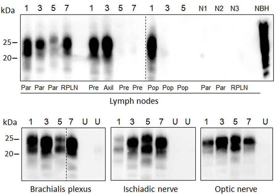
Figure 2. Protein misfolding cyclic amplification (PMCA) amplification of PrPSc (misfolded forms of the prion protein) in the lymphoreticular system and peripheral nerves of chronic wasting disease–affected animals from a study of prions in muscles of cervids with chronic wasting disease, Norway. We subjected lymph nodes and nerves collected from chronic wasting disease–affected cervids in Norway to serial PMCA before proteinase K digestion and Western blot analysis using Sha31 antibody. Example Western blots of samples from 1 representative animal of the different species and strains are shown; a more comprehensive result of all analyzed samples is summarized in Table 2. Lane designations: 1, reindeer A; 3, moose A; 5, moose C; 7, red deer. N lanes show lymph node from healthy, negative reindeer (N1), moose (N2), and red deer (N3). Dashed lines between images depict membrane splicing. Axil, axillary node; NBH, proteinase K–undigested bank vole brain homogenate used as electrophoretic migration marker of normal prion protein; par, parotid node; pre, prescapular node; pop, popliteal node; RPLN, retropharyngeal node; U, unseeded reaction included as a specificity control of PMCA reaction.
1These first authors contributed equally to this article.
2These authors are co–senior authors.