Volume 31, Number 9—September 2025
Research Letter
Characterization of Emerging Human Dirofilaria repens Infections, Estonia, 2023
Kalev Nõupuu, Maare Mõtsküla, Riina Pulges, Mikk Pauklin, and Urmas Saarma
Figure 1
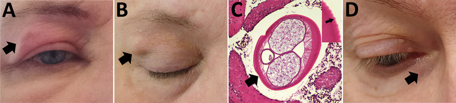
Figure 1. Autochthonous Dirofilaria repens infections in 2 women, Estonia, 2023. A–C) Case-patient 1, showing a painful subcutaneous lesion with edema on her right eyelid (A; black arrow). Two months later, the edema resolved, but a 1.5 cm subcutaneous nodule persisted (B; black arrow). Histopathologic examination of the removed nodule confirmed Dirofilaria spp. (C; larger black arrow); distinctive transverse striations on the cuticle with average distance of 12 µm between the peaks suggested D. repens (inset; small black arrow). Hematoxylin and eosin stain; original magnification ×100. The diameter of the parasite was 390 × 529 µm. D) Case-patient 2, showing a subcutaneous nodule under the right lower eyelid (black arrow).
Page created: July 09, 2025
Page updated: August 26, 2025
Page reviewed: August 26, 2025
The conclusions, findings, and opinions expressed by authors contributing to this journal do not necessarily reflect the official position of the U.S. Department of Health and Human Services, the Public Health Service, the Centers for Disease Control and Prevention, or the authors' affiliated institutions. Use of trade names is for identification only and does not imply endorsement by any of the groups named above.