Volume 31, Number 9—September 2025
Research Letter
Characterization of Emerging Human Dirofilaria repens Infections, Estonia, 2023
Cite This Article
Citation for Media
Abstract
Mosquitoborne diseases are a growing threat to public health worldwide. Human dirofilariasis, caused by the nematode Dirofilaria repens and transmitted by mosquitoes from various genera, has recently expanded into new areas of Europe. In this article, we report molecularly confirmed autochthonous human D. repens infections in Estonia.
Human dirofilariasis, caused by nematodes of the genus Dirofilaria, is a mosquitoborne parasitosis with growing public health importance. In Europe, the main causative species is D. repens, and infections with D. immitis are less frequent. Mosquitoes play a crucial role in the transmission of infectious larvae, and suitable species span various mosquito genera, including Aedes, Anopheles, and Culex (1). The definitive hosts of D. repens nematodes are domestic and wild carnivores. Humans are considered accidental hosts, in whom the parasitic larvae typically develop into a nonfertile stage. Although most human cases are subclinical, D. repens infection might occasionally result in subcutaneous swelling with subsequent development of subcutaneous nodular lesions. The clinical manifestations can include a mobile mass within the conjunctiva (2) and can lead to irreversible ocular damage (3). In rare cases, microfilaremia has been described (3,4). The distribution of D. repens nematodes includes countries in Europe, Africa, Middle East, Asia, and South America (1). In Europe, the parasite has recently spread north, and cases of human dirofilariasis caused by D. repens infection have emerged in Lithuania, Latvia, and Finland (5,6). D. repens nematodes have been reported in dogs in Estonia (7,8). Here, we describe 2 molecularly confirmed human D. repens infection cases from Estonia.
Figure 1
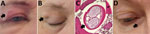
Figure 1. Autochthonous Dirofilaria repensinfections in 2 women, Estonia, 2023. A–C) Case-patient 1, showing a painful subcutaneous lesion with edema on her right eyelid (A; black arrow). Two months later,...
In February 2023, a 46-year-old woman in Estonia with no travel history abroad was referred to a neurology service for investigation of recurrent headache and painful facial subcutaneous nodular lesions with accompanying edema. The patient described a 1.5–2 cm palpable lesion that remained at the same location for 1–2 days before disappearing and reappearing elsewhere. Her symptoms persisted for 1 month, during which she observed nodules and edema on her upper (Figure 1, panel A) and lower eyelids, forehead, lips, and scalp. Two months later, a permanent 1.5 cm subcutaneous nodule developed in her right upper eyelid (Figure 1, panel B), and the patient was referred to an ophthalmologist. We surgically removed the nodule and sent it for histopathologic examination. The parasite in the nodule was confirmed as Dirofilaria spp. (Figure 1, panel C). After the removal of the parasite, her symptoms resolved and did not recur.
In February 2023, a 77-year-old woman in Estonia with no travel history abroad was referred to an ophthalmologist because of a nodular lesion under the right lower eyelid (Figure 1, panel D). The patient reported an episode of a mild, painful swelling that preceded the formation of the lesion. The oval lesion (1 × 1.5 cm) was located nasally under the right lower eyelid next to the orbital rim, was not painful and was freely movable, and was not adhered to the orbital rim or lacrimal sac. Initially, we recommended conservative observation; however, over the next 5 months, the lesion grew, and mild edema reappeared. Because of a clinical suspicion of a tumor, we surgically excised the lesion. The specimen was submitted for a histopathologic examination, which identified Dirofilaria spp.
No treatment guidelines for human D. repens infection have been established; however, surgical removal of the parasite is usually sufficient, and pharmacologic treatment is rarely necessary (3). After the removal of the encapsulated parasite in the 2 cases described, the symptoms resolved. No systemic treatment was applied. Additional data for both human cases are provided (Appendix).
Figure 2
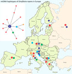
Figure 2. Distribution map of emerging Dirofilaria repenshaplotypes (n = 13) in Europe, including samples from Estonia, 2023. Haplotypes were derived from the median joining network analysis (left), illustrating relationships...
We conducted molecular genetic analysis for species identification and phylogenetic inference on samples collected from both patients (Appendix). We submitted sequences to GenBank (accession nos. PQ608665, PQ608666, and PQ608671). We conducted species identification on the basis of the 331 bp sequence by using homology search with nucleotide BLAST (https://blast.ncbi.nlm.nih.gov) and identified both pathogens as D. repens nematodes. On the basis of the longer fragment of cox1 (570 bp), we built a phylogenetic network that comprised the human isolate In2 from Estonia and 38 highly homologous sequences from GenBank (Figure 2). In that network, the human isolate In2 from Estonia formed a unique haplotype 1, suggesting low-level divergence from the central haplotype 2. Of note, D. repens is genetically closest to a newly described species, D. asiatica (9) (Appendix).
We report 2 autochthonous human D. repens infections in Estonia, highlighting the importance of recognizing this emerging threat. The geographic distribution of D. repens may have expanded because of climate change, enabling the parasite and its vectors to adapt in colder regions and spread the infection northward (3,8). Therefore, it is necessary to increase the awareness of the parasite among healthcare professionals working in Estonia. In both of these cases, the diagnosis was delayed, and several unnecessary tests were conducted because of a lack of knowledge about dirofilariasis. The common nodular lesions in the facial region might mimic tumors, granulomas, or cysts (10). Because no commercially available tests are available to diagnose D. repens infection from blood samples, molecular analysis of the parasite is essential for diagnosis (3).
Dr. Nõupuu is working as a senior ophthalmologist and eye surgeon in the Eye Clinic of Tartu University Hospital. His primary research interests are eye diseases, including those caused by zoonotic parasites.
Acknowledgments
We thank Mikk Tooming for his technical assistance and Sander Lupp for the histopathologic analysis.
This research was financed by the Estonian Ministry of Education and Research (grant nos. PRG1209 and TK215 to U.S.).
References
- Perles L, Dantas-Torres F, Krücken J, Morchón R, Walochnik J, Otranto D. Zoonotic dirofilariases: one, no one, or more than one parasite. Trends Parasitol. 2024;40:257–70. DOIPubMedGoogle Scholar
- Redón-Soriano M, Blasco A, Gomila B, González-Sánchez M, Simón F, Esteban JG. Subconjunctival human dirofilariasis by Dirofilaria repens in the Mediterranean Basin. Am J Ophthalmol Case Rep. 2022;26:
101570 . DOIPubMedGoogle Scholar - Capelli G, Genchi C, Baneth G, Bourdeau P, Brianti E, Cardoso L, et al. Recent advances on Dirofilaria repens in dogs and humans in Europe. Parasit Vectors. 2018;11:663. DOIPubMedGoogle Scholar
- Potters I, Vanfraechem G, Bottieau E. Dirofilaria repens Nematode infection with microfilaremia in traveler returning to Belgium from Senegal. Emerg Infect Dis. 2018;24:1761–3. DOIPubMedGoogle Scholar
- Pietikäinen R, Nordling S, Jokiranta S, Saari S, Heikkinen P, Gardiner C, et al. Dirofilaria repens transmission in southeastern Finland. Parasit Vectors. 2017;10:561. DOIPubMedGoogle Scholar
- Deksne G, Davidson RK, Buchmann K, Kärssin A, Kirjušina M, Gavarāne I, et al. Parasites in the changing world - Ten timely examples from the Nordic-Baltic region. Parasite Epidemiol Control. 2020;10:
e00150 . DOIPubMedGoogle Scholar - Jokelainen P, Mõtsküla PF, Heikkinen P, Ülevaino E, Oksanen A, Lassen B. Dirofilaria repens microfilaremia in three dogs in Estonia. Vector Borne Zoonotic Dis. 2016;16:136–8. DOIPubMedGoogle Scholar
- Alsarraf M, Levytska V, Mierzejewska EJ, Poliukhovych V, Rodo A, Alsarraf M, et al. Emerging risk of Dirofilaria spp. infection in Northeastern Europe: high prevalence of Dirofilaria repens in sled dog kennels from the Baltic countries. Sci Rep. 2021;11:1068. DOIPubMedGoogle Scholar
- Colella V, Young ND, Manzanell R, Atapattu U, Sumanam SB, Huggins LG, et al. Dirofilaria asiatica sp. nov. (Spirurida: Onchocercidae) - Defined using a combined morphological-molecular approach. Int J Parasitol. 2025;55:461–74. DOIPubMedGoogle Scholar
- Simón F, Siles-Lucas M, Morchón R, González-Miguel J, Mellado I, Carretón E, et al. Human and animal dirofilariasis: the emergence of a zoonotic mosaic. Clin Microbiol Rev. 2012;25:507–44. DOIPubMedGoogle Scholar
Figures
Cite This ArticleOriginal Publication Date: August 19, 2025
Table of Contents – Volume 31, Number 9—September 2025
| EID Search Options |
|---|
|
|
|
|
|
|
Please use the form below to submit correspondence to the authors or contact them at the following address:
Urmas Saarma, Department of Zoology, Institute of Ecology and Earth Sciences, University of Tartu, J. Liivi 2, 50409 Tartu, Estonia
Top