Skip directly to site content Skip directly to page options Skip directly to A-Z link Skip directly to A-Z link Skip directly to A-Z link

Disclaimer: Early release articles are not considered as final versions. Any changes will be reflected in the online version in the month the article is officially released.

Volume 32, Number 5—May 2026

Dispatch

Tropism and Replication Competence of Cattle Influenza A(H5N1) Genotype B3.13 Virus in Human Bronchus and Lung Tissue

Kenrie P.Y. Hui, John C.W. Ho, Ka-Chun Ng, Richard J. Webby, Malik Peiris, John M. Nicholls, and Michael C.W. ChanComments to Author 
Author affiliation: Centre for Immunology & Infection, Hong Kong, China (K.P.Y. Hui, J.C.W. Ho, M. Peiris, M.C.W. Chan); School of Public Health, Li Ka Shing Faculty of Medicine, University of Hong Kong, Hong Kong (K.P.Y. Hui, K.-C. Ng, M. Peiris, M.C.W. Chan); St. Jude Children’s Research Hospital, Memphis, Tennessee, USA (R.J. Webby); School of Clinical Medicine, Li Ka Shing Faculty of Medicine, University of Hong Kong, Hong Kong (J.M. Nicholls)

Main Article

Figure 2

Immunohistochemical stain of nucleoprotein from samples in a study of tropism and replication competence of cattle influenza A(H5N1) genotype B3.13 virus in human bronchus and lung tissue. Formalin-fixed, paraffin-embedded sections of human bronchus (A) and lung (B) tissue explants at 48 hours postinfection are shown. Cells positive for influenza A nucleoprotein are indicated by red-brown color. Green arrows indicate ciliated cells; yellow arrows indicate nonciliated cells. Images are representatives of 3 separate donors. Cattle influenza A(H5N1) genotype B.13 virus strains A/dairy_cow/Ohio/B24OSU-439/2024 (H5N1/439) and A/dairy_cow/Texas/98638/2024 (H5N1/98638) are compared with historical human isolates of highly pathogenic avian influenza strains H5N1/483, H5N6/39715, and H1N1pdm/415742 (Appendix Tables 1, 2). Scale bars indicate 100 μm.

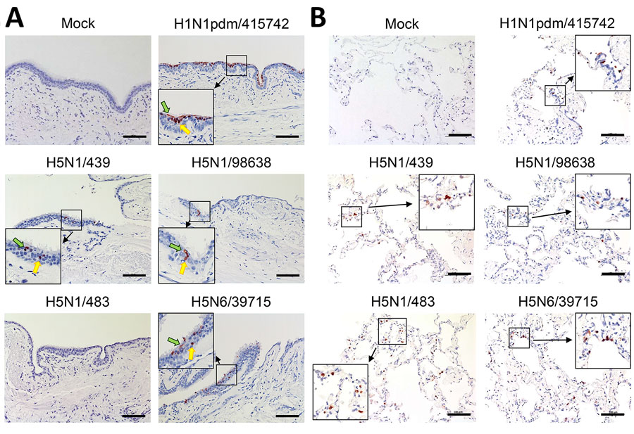
Figure 2. Immunohistochemical stain of nucleoprotein from samples in a study of tropism and replication competence of cattle influenza A(H5N1) genotype B3.13 virus in human bronchus and lung tissue. Formalin-fixed, paraffin-embedded sections of human bronchus (A) and lung (B) tissue explants at 48 hours postinfection are shown. Cells positive for influenza A nucleoprotein are indicated by red-brown color. Green arrows indicate ciliated cells; yellow arrows indicate nonciliated cells. Images are representatives of 3 separate donors. Cattle influenza A(H5N1) genotype B.13 virus strains A/dairy_cow/Ohio/B24OSU-439/2024 (H5N1/439) and A/dairy_cow/Texas/98638/2024 (H5N1/98638) are compared with historical human isolates of highly pathogenic avian influenza strains H5N1/483, H5N6/39715, and H1N1pdm/415742 (Appendix Tables 1, 2). Scale bars indicate 100 μm.

Main Article

Page created: March 24, 2026
Page updated: April 01, 2026
Page reviewed: April 01, 2026
The conclusions, findings, and opinions expressed by authors contributing to this journal do not necessarily reflect the official position of the U.S. Department of Health and Human Services, the Public Health Service, the Centers for Disease Control and Prevention, or the authors' affiliated institutions. Use of trade names is for identification only and does not imply endorsement by any of the groups named above.
file_external