Three Fatal Gestational Psittacosis Cases Caused by Chlamydia psittaci Strains Belonging to Closely Related Lineages, Japan
Atsuko Nishino, Yukiko Nakura, Yukiko Sassa-O’Brien, Momoko Soeda, Hirokazu Sugii, Kanako Shimizu, Shiro Miura, Yumiko Sato, Michinobu Yoshimura
1, Michiko Kodama, and Itaru Yanagihara

Author affiliation: Research Institute, Osaka Women’s and Children’s Hospital, Osaka, Japan (A. Nishino, Y. Nakura, M. Yoshimura, I. Yanagihara); The University of Osaka, Osaka (A. Nishino, M. Kodama, I. Yanagihara); Tokyo University of Agriculture and Technology, Tokyo, Japan (Y. Sassa-O’Brien); NHO Nagasaki Medical Center, Nagasaki, Japan (M. Soeda, S. Miura); NHO Iwakuni Clinical Center, Yamaguchi, Japan (H. Sugii, Y. Sato); Tannan Health Welfare Center, Fukui, Japan (K. Shimizu); Maizuru Kyosai Hospital, Kyoto, Japan (K. Shimizu)
Main Article
Figure 5
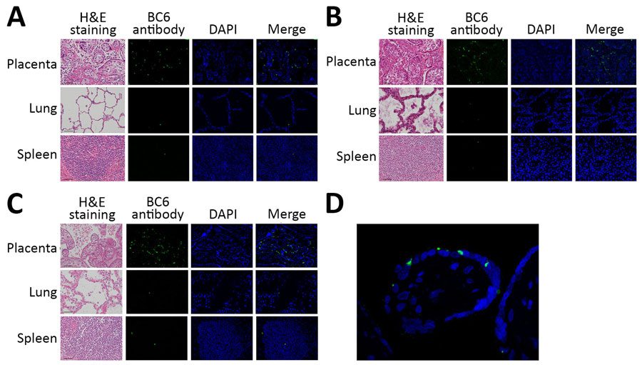
Figure 5. Histologic findings from 3 fatal gestational psittacosis cases caused by C. psittaci strains belonging to closely related multilocus sequence typing lineages, Japan, 2017–2024. A–C) Hematoxylin and eosin staining (scale bar = 50 µm; original magnification ×20) and immunofluorescence microscopy of the placenta, lung, and spleen are shown for case FO-01 (A), case YO-02 (B), and case NO-03 (C). Specific fluorescence observed via immunofluorescence using C. psittaci BC6 rabbit antibody staining. Nuclei were stained with DAPI. Merge column indicates BC6 antibody staining and DAPI. Original magnification ×20. D) High-magnification view (original magnification ×60) of the villi. DAPI, 4′,6-diamidino-2-phenylindole; H&E, hematoxylin and eosin.
Main Article
Page created: April 01, 2026
Page updated: May 08, 2026
Page reviewed: May 08, 2026
The conclusions, findings, and opinions expressed by authors contributing to this journal do not necessarily reflect the official position of the U.S. Department of Health and Human Services, the Public Health Service, the Centers for Disease Control and Prevention, or the authors' affiliated institutions. Use of trade names is for identification only and does not imply endorsement by any of the groups named above.