Volume 32, Number 6—June 2026
Research
Role of Households with Children in Community Spread of Multidrug-Resistant Enterobacterales, St. Louis, Missouri, USA
Cite This Article
Citation for Media
Abstract
Community-acquired multidrug-resistant (MDR) Enterobacterales bacteria are an increasing public health concern, yet whether households play a role in community spread remains unclear. We investigated 150 households with children in St. Louis, Missouri, USA, for MDR Enterobacterales. We cultured swab specimens from household members and environmental surfaces for identification and antimicrobial susceptibility testing. We also performed whole-genome sequencing in the 53 (35%) households where >1 MDR Enterobacterales species were recovered. Enterobacter hormaechei predominated, followed by Klebsiella pneumoniae and Pantoea species. Whole-genome sequencing revealed closely related strains shared between persons and environmental surfaces, suggesting potential intra-household transmission. We identified >1 horizontal gene transfer event between Enterobacterales genera within a household. On multivariable analysis, households that had children attending daycare, a member with an ADHD diagnosis, and dog ownership were associated with increased odds of household MDR Enterobacterales colonization. Households likely serve as major contributors in acquisition and community spread of MDR Enterobacterales.
Multidrug resistant (MDR) Enterobacterales bacteria have emerged as a serious public health threat. Of major concern is the increasing incidence of extended-spectrum cephalosporin resistance (ESCR) in Enterobacterales. This resistance pattern is commonly associated with the production of extended-spectrum β-lactamases (ESBL), antimicrobial resistance genes (ARGs) carried on mobile genetic elements (MGEs) such as plasmids, which often carry other ARGs. The incidence of infections has increased dramatically during the past decade, reaching nearly 200,000 infections and >9,000 deaths per year in the United States (1). Although ESBL Enterobacterales were once largely healthcare-associated pathogens, during the past 2 decades, most ESBL Enterobacterales infections have been caused by clinically and genetically distinct strains that have emerged in the community (2).
Adding to the complexity of this group of pathogens, the determinants of resistance yielding the ESCR phenotype might be chromosomal, transferable ARGs on MGEs, or contain both, and are often MDR. Invasive infections caused by community- acquired ESCR Enterobacterales strains are increasingly being reported in young children and persons without major healthcare exposures (3,4). Of note, those strains are resistant to antimicrobial drugs that are uncommonly used in children; therefore, overuse is unlikely to be driving this resistance.
Factors promoting MDR and ESCR Enterobacterales acquisition and infection in the community are largely unknown. However, in households with an adult known to have acquired ESBL Enterobacterales from healthcare exposures, transmission incidence has been described as upwards of 67% (5,6). Furthermore, in a multicenter investigation of MDR (primarily ESBL) Enterobacterales in children from Chicago, Illinois, USA, we found that MDR Enterobacterales acquisition reflected geographic clustering and lacked association with the factors driving primary acquisition in adults (e.g., antimicrobial drug and healthcare exposures) (7,8). We also found that, compared with antimicrobial-sensitive Enterobacterales infections in children, blaCTX-M-9-type ESBL Enterobacterales infections were nearly 5 times more likely to be community-acquired (8). To devise strategies to prevent community-acquired MDR Enterobacterales infections, we must first understand key reservoirs for acquisition, including sources of transmission outside of healthcare settings, the role of the natural and built environment in pathogen transmission, and epidemiologic factors associated with community-acquired MDR Enterobacterales acquisition, transmission, and infection.
We believe households are major drivers of MDR Enterobacterales spread in the community and that environmental surfaces are major reservoirs of MDR Enterobacterales in households. That hypothesis is supported by observations that MDR Enterobacterales strains can persist on surfaces in healthcare environments for up to 30 months, likely contributing to healthcare-associated transmission (9–11). Relevant to those findings, prior studies of household transmission of community-acquired methicillin-resistant Staphylococcus aureus (MRSA) in St. Louis, Missouri, USA, have demonstrated that multiple environmental surfaces serve as reservoirs for community-acquired MRSA transmission (12–15). In addition, households with a higher burden of environmental community-acquired MRSA contamination were 4-fold more likely to enable transmission (15). In addition, we believe that critical epidemiologic risk factors for colonization, including the presence of preschool-age children who attend daycare (16) and having a pet dog in the household, would be associated with MDR Enterobacterales colonization (17).
We used a biorepository of human and household surface samples and associated epidemiologic metadata collected from households of pediatric and adult community participants to understand the clinical and molecular epidemiology of community-acquired MDR Enterobacterales, with a focus on ESCR Enterobacterales. We highlight the household whole-genome sequencing (WGS) data for Enterobacter spp., a well-known cause of healthcare-associated infections, which might represent an underrecognized source of community acquisition of antimicrobial drug resistance.
Study Settings and Population
We used a biorepository and detailed epidemiologic metadata from a well-curated population of 150 otherwise healthy children with community-acquired S. aureus infections and their household contacts (n = 489) enrolled in the SHINE study from 2015–2021 in metropolitan St. Louis, Missouri, USA (Clinicaltrials.gov, identification no. NCT02572791). Through the SHINE study, research visits were conducted in participants’ homes. Children were defined as 0–18.99 years old. Detailed clinical and epidemiologic data were collected from each participant, including demographics, medical history, topical and systemic antimicrobial drug use, activities outside of the home, personal hygiene practices, and interactions between household contacts. Detailed information regarding household characteristics were also collected, including household environmental cleaning practices, renting versus owning, number of bedrooms, and the presence and characteristics of pets. All data were collected prospectively through the SHINE study and were entered into REDCap, a secure, HIPAA-compliant, web-based data application (18). At each study visit, colonization samples were collected from the anterior nares, axillae, and inguinal folds by using BD Eswabs (BD, https://www.bd.com). Rectal or perirectal swab specimens were not collected.
Through the SHINE study, samples were collected from up to 21 household environmental surfaces. A 250-µL aliquot of liquid amies transport media from each swab was enriched and suspended in tryptic soy broth plus 20% glycerol and frozen at −80°C. For this study, we used samples from households collected at SHINE study enrollment (natural history phase). The Washington University Institutional Review Board approved study procedures. Informed consent was obtained for all participating household members.
Bacterial Isolates and Antimicrobial Susceptibility Testing
We chose to use inguinal fold swab specimens on the basis of a higher likelihood of colonization because of proximity to the rectum. We transferred swab suspension samples and environmental surface samples to tryptic soy broth and incubated them overnight at 37°C. We used a 1-μL inoculation loop to streak the enriched broth onto membrane fecal coliform agar to evaluate the presence of Enterobacterales bacteria. In addition, we spread-plated 100 μL of the broth onto CHROMagar ESBL media (CHROMagar, https://www.chromagar.com) (19). We incubated plates at 37°C for 16–20 hours, subcultured the resulting colonies on CHROMagar, and incubated them overnight at 37°C. After incubation, we resuspended the colonies in saline and adjusted to a 0.5–0.63 McFarland standard for isolate identification and antimicrobial susceptibility testing using the automated VITEK 2 system (bioMérieux, https://www.biomerieux.com), according to the manufacturer’s instructions.
We used a broad definition for MDR Enterobacterales because isolates were ESCR, with some demonstrating resistance to carbapenems, all of which were resistant to >2 antimicrobial classes. We selected the colonies we defined as MDR Enterobacterales for further testing (20). We chose a subset of the resistant isolates for WGS if they were from households where Enterobacterales were recovered from >1 surface or person. We extracted DNA by using the QIAGEN DNeasy Blood and Tissue Kit (QIAGEN, https://www.qiagen.com) according to the manufacturer’s instructions and shipped the extracted genomic DNA to the Rush University Medical Center Genomics and Microbiome core facility (Chicago, Illinois, USA) for WGS.
WGS
We performed bacterial WGS by using standard shotgun sequencing methods, as described previously (19). In brief, we prepared genomic DNA for sequencing by using a NEXTFLEX rapid XP DNA sequencing kit (Revvity, https://www.revvity.com) implemented on a Sciclone G3 NGSx iQ (Revvity) workstation. We normalized DNA inputs to 10 ng and used 10 cycles of amplification. After magnetic bead cleanup (0.8× ratio of beads to template, vol:vol), we sequenced libraries by using an Illumina NovaSeq X (Illumina, https://www.illumina.com) instrument using a 10 billion cluster flowcell lane. Libraries were created by the Rush University Medical Center for Genomics and Microbiome core facility, and sequencing was performed by the DNA Services Core, Carver Biotechnology Center, University of Illinois Urbana-Champaign (Urbana-Champaign, Illinois, USA).
Bioinformatic Analysis
FASTQ files underwent QC screening, and we assembled and analyzed them by using the Bactopia pipeline (21). In brief, we only further analyzed FASTQ files if they met the following parameters: estimated genome coverage >20×, mean per-read quality score >Q12, mean post-trimming read length >49 bp, and <500 total contigs. We quality filtered Illumina reads by using Trimmomatic (22) and assembled de novo by using SPAdes (23). We used Prodigal (24) to predict gene sequences and annotated them with Prokka (25). We assessed antimicrobial resistance content by using AMRFinder Plus (26). We defined core genes by using Roary (27). We generated a phylogenetic tree on the basis of a core gene alignment by using IQtree (28). We generated a maximum-likelihood tree by running 1,000 bootstrap replicates under the generalized time-reversible model of evolution. We inferred the maximum-likelihood phylogeny from the core genome alignment by using IQ-TREE under the Hasegawa–Kishino–Yano nucleotide substitution model with 1,000 ultrafast bootstrap replicates and 1,000 SH-like approximate likelihood ratio test replicates. We visualized and annotated the tree by using iTOL version 4 (29). We calculated the core genome pairwise single-nucleotide polymorphism distance for each sample with snp-dists (30) and completed pangenome wide comparison of genomes by using Scoary (31). We reconstructed, typed, and clustered plasmids by using MOB-suite (32). We performed clustering for plasmids with a mash distance <0.05 with >85% similarity in length. We considered plasmid transfer if a mobilizable or conjugative plasmid within the same primary cluster was detected in a different species within the same household. Sequence data are available in the National Center for Biotechnology Information Sequence Read Archive (https://www.ncbi.nlm.nih.gov/sra; BioProject no. PRJNA1257399).
Statistical Analysis
We used a retrospective case–control study design to assess factors associated with household colonization with MDR Enterobacterales. We presented the descriptives numerical variable by mean +SD and of categorical variables by counts and percentages (33). We conducted bivariate analysis by using a 2-sample t-test and χ2 test for independence. We conducted multivariate analysis of the binary outcome by using logistic regression. We selected covariates in the final logistic regression by the LASSO method (34,35) first, and then by clinical importance of the variables because of a large number of covariates associated with households and household members and pets (12,36).
We performed logistic regression analysis to identify significant household clinical and nonclinical factors associated with household colonization with MDR Enterobacterales. On the basis of the observation numbers, robustness to reporting, prevarication bias, and biologic plausibility regarding Enterobacterales carriage, we selected a covariate with a p<0.25 in univariate analysis for inclusion in a multivariable model by using a manual forward selection approach in which variables having a p = 0.10 remained in the model. We addressed potential confounding effects by retaining variables whose exclusion from the models changed the effect of the other covariates by >10%. We tested interactions between independent variables and expanded the final multivariable models to include the significant (p<0.10) interaction terms. We checked for any collinearities between independent variables before multivariable analysis and made selections between collinear variables on the basis of an improved model fit as shown by the Akaike information criterion and Bayesian information criterion (36,37). We expressed bivariate and multivariate associations as odd ratios (ORs) and corresponding 95% CIs. Statistical significance was indicated by p<0.05.
Characteristics of Households in the Study Population
We analyzed 150 households and their characteristics. The mean age of the 639 participants was 20.86 (SD +6.30); for race, 73% identified as White, 24% as Black, and 3% as mixed race or Asian descent. In addition, >1 household member held a college degree (43%), private insurance (77%) or Medicaid insurance (31%). All households had >1 child (52% of all participants were children), 71% had >1 child in daycare, 51% had >1 dog, and the average number of residents per household was 4.31 (SD +1.34).
We univariately analyzed ≈100 variables from clinical and epidemiologic data. Of those, we summarized 20 risk factors from the univariate analysis results along with their bivariate associations on the basis of the presence and absence of MDR Enterobacterales (Table 1).
Characteristics of Bacterial Isolates at the Household Level
We tested 3,201 samples from 627 humans and 2,574 surfaces in 150 households. Enterobacterales strains phenotypically identified as MDR had been recovered from 53 (35%) of 150 households. Of the 120 MDR Enterobacterales isolated from 53 households, most were Enterobacter spp. (71%, n = 85), Pantoea spp. (12%, n = 14), and Klebsiella spp. (8%, n = 10). The household surfaces most commonly harboring MDR Enterobacterales were the kitchen sink faucet handle (20.7%), sofa (9%), bedsheets (6%), oven door handle (6%), and refrigerator door handle (3%). MDR Enterobacterales were identified in the inguinal folds of 25 (4%) household members.
WGS Analysis of MDR Enterobacterales strains
We chose a subset of isolates for WGS if they were recovered from households where MDR Enterobacterales were found on >1 household surface or household member. Of the 94 samples sequenced, 93 passed pipeline QC metrics. Of those, 76 were Enterobacterales on the basis of genome taxonomy database toolkit taxonomic classification (Table 2). The most common species detected were members of the Enterobacter cloacae complex (most were E. hormaechei [N = 47]), followed by K. pneumoniae (N = 10). All isolates were ESCR; 10 were also resistant to >1 carbapenem, none of which were found to contain a transmissible carbapenemase gene.
We conducted a relatedness analysis for all species or sequence types (STs) with >4 isolates (K. pneumoniae [n = 10], S. marcescens [n = 4], E. hormaechei ST50 [n = 5] and ST108 [n = 5]). This analysis revealed clustering of isolates within the same households from multiple surfaces and household members (Appendix Table 1).
We reconstructed, typed, and clustered plasmid sequences to assess whether plasmids with similar identity were found across different species isolated from the same household. A total of 251 plasmids were reconstructed across 69 genomes into 100 unique clusters. Across those clusters, we detected >1 potential plasmid transfer event. A plasmid with the same conjugative relaxase type and an identical mash distance to the nearest MOB-suite database reference (GenBank accession no. CP032172) was detected in a K. pneumoniae isolate recovered from the inguinal fold of a participant child and Proteus mirabilis isolate recovered from the inguinal fold of the child’s mother.
Figure
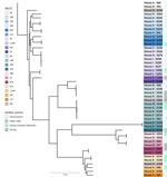
Figure. Phylogenetic tree based on core gene alignment for isolated Enterobacter hormaecheispecies (n = 47) in study of the role of households with children in community spread of multidrug-resistant...
Further analysis of the recovered E. hormaechei strains revealed a diversity of STs, with 23 unique STs detected (Figure; Appendix Table 2, Figure). Only 2 major STs (defined as >5 isolate per ST) were detected: ST50 (n = 5 isolates) and ST108 (n = 5 isolates). The remaining 47 were distributed across minor STs (n < 5 isolates per ST). All isolates had a blaACT variant ampC gene detected. Among the 47 isolates, 15 were detected from a human source (3 from inguinal folds, 12 from bedsheets) and 32 from an environmental source. ST distribution, AMR gene count, and plasmid count was similar among environmental and human isolates (Appendix Table 2). To assess differential gene content between environmental and human isolates, we conducted a pangenome analysis. However, no candidate genes remained statistically significant after Benjamini-Hochberg correction (false discovery rate <0.05). K. pneumoniae, the second largest group of strains assessed by WGS revealed primarily blaSHV-ESBL and most were also MDR (Table 3).
Analysis of Factors Associated with MDR Enterobacterales
The 53 households with MDR Enterobacterales (cases) were compared to 97 households without MDR Enterobacterales (controls). Although 94 of 150 households identified having >1 family member with >1 health conditions (Table 1; Appendix Table 3), none of those conditions were found to be associated with MDR Enterobacterales colonization on bivariate analysis; primary conditions reported were often mild or common such as asthma and seasonal allergies. However, households reporting >1 antimicrobial drug prescription in the previous 12 months were more common in controls than cases and was inversely associated with MDR Enterobacterales colonization (OR = 0.48, 95% CI 0.24–0.96; p = 0.04). Having smaller homes, fewer rooms, and lower square feet per person were positively associated with MDR Enterobacterales colonization on bivariate analysis.
In the final multivariable logistic regression model (Table 4), factors found to be associated with a lower likelihood of MDR Enterobacterales colonization were households identifying as predominantly White race (adjusted OR [aOR] = 0.18, 95% CI 0.06–0.49; p<0.01) and having ≥1 member of the family with private insurance trended toward significance and was included in the final model (aOR = 0.44, 95% CI 0.16–1.22; p = 0.11). Factors associated with increased risk for household colonization with MDR Enterobacterales included having >1 with a diagnosis of ADHD (aOR = 3.47, 95% CI 1.34–9.41; p = 0.01), >1 minor attending daycare (aOR = 2.86, 95% CI 1.07–8.38, p = 0.04), and >1 dog (aOR = 3.31, 95% CI 1.27–3.31; p = 0.02). Similar differences can be found between aOR and OR (Appendix Table 4).
In this study, we focused on understanding the role of the household in acquisition and transmission of MDR Enterobacterales, factors associated with increased or decreased risk for household colonization, the principal genetic determinants, and the relatedness of MDR Enterobacterales strains in community settings. Our research program investigates antimicrobial drug resistance in the community through a One Health lens (38).
Enterobacterales that exhibit higher level resistance are designated as high priority in the 2024 WHO priority report (39). Of note, studying colonizing isolates overcomes biases that are intrinsic to surveillance systems reliant on passively collected clinical isolates (40), which is critical because colonization frequently precedes infection, and asymptomatic carriers can serve as sources of onward MDR organism transmission. In particular, colonization with MDR and ESCR Enterobacterales in children can last months or years, and silent dissemination of transmissible ARGs in Enterobacterales has been described in healthy pediatric populations (41–43).
We found that, in households not known to previously harbor MDR Enterobacterales, the prevalence in midwestern US communities was high, 35%. The average household size was 4.31, and 100% of households had >1 child. Those findings are consistent with the continued increases of ESCR Enterobacterales in community settings, despite the successes of aggressive infection prevention and control campaigns in healthcare settings (3,4). We also found the presence of other major transmissible ARGs, such as fluoroquinolone, sulfonamide, fosfomycin, and tetracycline resistance genes, along with the presence of multiple conjugative plasmids among isolates (Table 3; Appendix Table 2). Previous studies that have investigated household transmission of MDR Enterobacterales were predominately among previously hospitalized adult patients with known colonization (6).
Although ESBL-producing K. pneumoniae and P. mirabilis were recovered from households, we did not find a major presence of ESBL-producing E. coli. However, we did find the presence of high-risk E. coli clones (e.g., ST131, ST69, ST127, ST73) known for their epidemic potential and high potential for acquiring or having ARGs and MGEs.
Of note, the Enterobacter cloacae complex group of bacteria are ubiquitous in nature; however, these bacteria are most found in healthcare-associated infections in hospitalized patients or persons with antimicrobial or healthcare exposures. We were surprised to see such a high level of colonization in relatively healthy community households. In addition, most of our isolates within Enterobacter cloacae complex were E. hormaechei, which is often MDR and known to cause extraintestinal healthcare-associated infections, (e.g., urinary tract, bloodstream, and pneumonia), and can persist in healthcare environments (44). We found evidence of clustering of E. hormaechei within and between households, suggesting that household and community reservoirs might be a major source of community acquisition and spread of these pathogens.
The first limitation of our study is the relatively small sample size and large number of variables. We could not study the nonlinear and nonmonotonic effects of several count variables, and the suggested multivariate model might not be optimal for the given data. However, the relatively large prevalence of Enterobacterales has shown good statistical classification power of the study population. The multivariate model with 5 covariates showed good predictive power with 81.2% area under the curve. The model classified households with 75% sensitivity and 75% specificity when the threshold was 0.35. Second, because of the relatively small sample size of non-White households, we cannot provide details about the effect modification of race and suspect that race might represent a proxy for differences in socioeconomic status in the region of study. Although we show the link between pets, in particular dog ownership, and the diagnosis of ADHD and their association with household colonization with MDR Enterobacterales, many non-White households, predominantly Black, did not have pets or family members with ADHD diagnosis, limiting the ability for further analysis of those variables. Third, the parent study of the biorepository used for this analysis was initially designed to assess S. aureus household colonization, and households were selected on the basis of having a healthy child who had an S. aureus skin and soft tissue infection. Therefore, inguinal swab specimens were used to assess for household member colonization for MDR Enterobacterales because rectal or perirectal swab specimens were not collected in the parent study. Although ideally rectal or perirectal swab specimens would have been used, prior surveillance studies have demonstrated that the main reservoir for Enterobacterales is the gastrointestinal tract, and the inguinal folds are the most colonized skin site outside of the perirectal area because of the proximity to the rectum (45–49). A military study demonstrated that the inguinal folds were the most sensitive anatomic site for detecting MDR gram-negative colonization outside of the perirectum (negative predictive value 98%–100% for ESBL Enterobacterales) (46). The inguinal folds in young and diapered children also have a high burden of colonization and secondary infection with bacteria because of incontinence, excess moisture, and friction (50). Within our study population, most households (123 of 150) had children 0–5 years of age (4,16). In addition, our finding of a 4% inguinal fold colonization rate is consistent with prior US-based pediatric studies of intestinal colonization with ESCR (4.4%) and ESBL-producing (3.5%) Enterobacterales in healthy US children collected during well child clinic visits (42). Finally, whereas we can demonstrate clustering within households and communities, our retrospective analysis of a single time point cannot establish timing nor directionality of transmission within or between households, surfaces, and its members.
In conclusion, households might serve as a major contributor to the acquisition and spread of MDR Enterobacterales in the community. Factors associated with household colonization with MDR Enterobacterales include having a pet dog or children who attend daycare. Our current and future prospective One Health focused studies continue to investigate community reservoirs of MDR Enterobacterales in humans, animals, the household, and the natural environment. Our study emphasizes the necessity of investigating community reservoirs and spread of MDR Enterobacterales to learn more about how to mitigate potential sources associated with colonization and infection in the community.
Mr. Breeze is a lead research specialist in the Department of Pediatrics, Division of Pediatric Infectious Diseases, at Emory University School of Medicine. His research interests include molecular identification of antimicrobial resistance genes in human, animal, and environmental populations and understanding the role of the environment in the spread of antibiotic-resistant bacteria in the community. Dr. Babiker is an assistant professor in the Department of Internal Medicine, Division of Infectious Diseases, at Rush Medical College. His research interests include the clinical and molecular epidemiology antimicrobial resistance organisms, the surveillance of antimicrobial resistance in low- and middle-income settings, and the role of the gastrointestinal microbiome in colonization resistance.
Acknowledgments
We thank the team of curators of the whole-genome MLST databases and National Institutes of Health GenBank database for curating the data and making them publicly available at https://www.ncbi.nlm.nih.gov/bioproject/PRJNA1257399. Sequence data is deposited in the Sequence Read Archive (accession nos. SRR33388617–95).
L.K.L was supported in part by the National Institute of Allergy and Infectious Diseases, National Institutes of Health (grant no. R21AI173471), the Marcus Foundation, and by the Emory University School of Medicine MP3 Initiative Award Program. A.B. was supported in part by an Antibacterial Resistance Leadership Group Early Faculty Seedling Award (award no. UM1AI104681).
References
- Centers for Disease Control and Prevention. Antibiotic resistance threats in the United States, 2019. 2019 [cited 2025 Dec 8]. https://www.cdc.gov/antimicrobial-resistance/media/pdfs/2019-ar-threats-report-508.pdf
- Price LB, Johnson JR, Aziz M, Clabots C, Johnston B, Tchesnokova V, et al. The epidemic of extended-spectrum-β-lactamase-producing Escherichia coli ST131 is driven by a single highly pathogenic subclone, H30-Rx. MBio. 2013;4:e00377–13. DOIPubMedGoogle Scholar
- Logan LK, Braykov NP, Weinstein RA, Laxminarayan R; CDC Epicenters Prevention Program. Extended-spectrum β-lactamase-producing and third-generation cephalosporin-resistant Enterobacteriaceae in children: trends in the United States, 1999–2011. J Pediatric Infect Dis Soc. 2014;3:320–8. DOIPubMedGoogle Scholar
- Lukac PJ, Bonomo RA, Logan LK. Extended-spectrum β-lactamase-producing Enterobacteriaceae in children: old foe, emerging threat. Clin Infect Dis. 2015;60:1389–97. DOIPubMedGoogle Scholar
- Haverkate MR, Platteel TN, Fluit AC, Cohen Stuart JW, Leverstein-van Hall MA, Thijsen SFT, et al. Quantifying within-household transmission of extended-spectrum β-lactamase-producing bacteria. Clin Microbiol Infect. 2017;23:46.e1–7. DOIPubMedGoogle Scholar
- Hilty M, Betsch BY, Bögli-Stuber K, Heiniger N, Stadler M, Küffer M, et al. Transmission dynamics of extended-spectrum β-lactamase–producing Enterobacteriaceae in the tertiary care hospital and the household setting. Clin Infect Dis. 2012;55:967–75. DOIPubMedGoogle Scholar
- Logan LK, Medernach RL, Rispens JR, Marshall SH, Hujer AM, Domitrovic TN, et al. Community origins and regional differences highlight risk of plasmid-mediated fluoroquinolone-resistant Enterobacteriaceae infections in children. Pediatr Infect Dis J. 2019;38:595–9. DOIPubMedGoogle Scholar
- Logan LK, Medernach RL, Domitrovic TN, Rispens JR, Hujer AM, Qureshi NK, et al. Clinical and molecular epidemiology of CTX-M-9-group-producing Enterobacteriaceae infections in children. Infect Dis Ther. 2019;8:243–54. DOIPubMedGoogle Scholar
- Freeman JT, Nimmo J, Gregory E, Tiong A, De Almeida M, McAuliffe GN, et al. Predictors of hospital surface contamination with extended-spectrum β-lactamase-producing Escherichia coli and Klebsiella pneumoniae: patient and organism factors. Antimicrob Resist Infect Control. 2014;3:5. DOIPubMedGoogle Scholar
- Kramer A, Schwebke I, Kampf G. How long do nosocomial pathogens persist on inanimate surfaces? A systematic review. BMC Infect Dis. 2006;6:130. DOIPubMedGoogle Scholar
- Thurlow CJ, Prabaker K, Lin MY, Lolans K, Weinstein RA, Hayden MK; Centers for Disease Control and Prevention Epicenters Program. Anatomic sites of patient colonization and environmental contamination with Klebsiella pneumoniae carbapenemase–producing Enterobacteriaceae at long-term acute care hospitals. Infect Control Hosp Epidemiol. 2013;34:56–61. DOIPubMedGoogle Scholar
- Hogan PG, Mork RL, Boyle MG, Muenks CE, Morelli JJ, Thompson RM, et al. Interplay of personal, pet, and environmental colonization in households affected by community-associated methicillin-resistant Staphylococcus aureus. J Infect. 2019;78:200–7. DOIPubMedGoogle Scholar
- Mork RL, Hogan PG, Muenks CE, Boyle MG, Thompson RM, Morelli JJ, et al. Comprehensive modeling reveals proximity, seasonality, and hygiene practices as key determinants of MRSA colonization in exposed households. Pediatr Res. 2018;84:668–76. DOIPubMedGoogle Scholar
- Fritz SA, Hogan PG, Singh LN, Thompson RM, Wallace MA, Whitney K, et al. Contamination of environmental surfaces with Staphylococcus aureus in households with children infected with methicillin-resistant S aureus. JAMA Pediatr. 2014;168:1030–8. DOIPubMedGoogle Scholar
- Mork RL, Hogan PG, Muenks CE, et al. Longitudinal, strain-specific Staphylococcus aureus introduction and transmission events in households of children with community-associated MRSA skin and soft-tissue infection: a prospective cohort study. Lancet Infect Dis. 2020;20:188–98. DOIPubMedGoogle Scholar
- Kaarme J, Riedel H, Schaal W, Yin H, Nevéus T, Melhus Å. Rapid increase in carriage rates of Enterobacteriaceae producing extended-spectrum β-lactamases in healthy preschool children, Sweden. Emerg Infect Dis. 2018;24:1874–81. DOIPubMedGoogle Scholar
- van den Bunt G, Fluit AC, Spaninks MP, Timmerman AJ, Geurts Y, Kant A, et al. Faecal carriage, risk factors, acquisition and persistence of ESBL-producing Enterobacteriaceae in dogs and cats and co-carriage with humans belonging to the same household. J Antimicrob Chemother. 2020;75:342–50. DOIPubMedGoogle Scholar
- Harris PA, Taylor R, Thielke R, Payne J, Gonzalez N, Conde JG. Research electronic data capture (REDCap)—a metadata-driven methodology and workflow process for providing translational research informatics support. J Biomed Inform. 2009;42:377–81. DOIPubMedGoogle Scholar
- Perry JD. A decade of development of chromogenic culture media for clinical microbiology in an era of molecular diagnostics. Clin Microbiol Rev. 2017;30:449–79. DOIPubMedGoogle Scholar
- Logan LK, Hujer AM, Marshall SH, Domitrovic TN, Rudin SD, Zheng X, et al. Analysis of β-lactamase resistance determinants in Enterobacteriaceae from Chicago children: a multicenter survey. Antimicrob Agents Chemother. 2016;60:3462–9. DOIPubMedGoogle Scholar
- Petit RA III, Read TD. Bactopia: a flexible pipeline for complete analysis of bacterial genomes. mSystems. 2020;5:e00190–20. DOIPubMedGoogle Scholar
- Bolger AM, Lohse M, Usadel B. Trimmomatic: a flexible trimmer for Illumina sequence data. Bioinformatics. 2014;30:2114–20. DOIPubMedGoogle Scholar
- Prjibelski A, Antipov D, Meleshko D, Lapidus A, Korobeynikov A. Using SPAdes de novo assembler. Curr Protoc Bioinformatics. 2020;70:
e102 . DOIPubMedGoogle Scholar - Hyatt D, Chen GL, Locascio PF, Land ML, Larimer FW, Hauser LJ. Prodigal: prokaryotic gene recognition and translation initiation site identification. BMC Bioinformatics. 2010;11:119. DOIPubMedGoogle Scholar
- Seemann T. Prokka: rapid prokaryotic genome annotation. Bioinformatics. 2014;30:2068–9. DOIPubMedGoogle Scholar
- Feldgarden M, Brover V, Haft DH, Prasad AB, Slotta DJ, Tolstoy I, et al. Validating the AMRFinder tool and resistance gene database using antimicrobial resistance genotype–phenotype correlations. Antimicrob Agents Chemother. 2019;63:e00483–19. DOIPubMedGoogle Scholar
- Page AJ, Cummins CA, Hunt M, Wong VK, Reuter S, Holden MT, et al. Roary: rapid large-scale prokaryote pan genome analysis. Bioinformatics. 2015;31:3691–3. DOIPubMedGoogle Scholar
- Nguyen LT, Schmidt HA, von Haeseler A, Minh BQ. IQ-TREE: a fast and effective stochastic algorithm for estimating maximum-likelihood phylogenies. Mol Biol Evol. 2015;32:268–74. DOIPubMedGoogle Scholar
- Letunic I, Bork P. Interactive tree of life (iTOL) v5: an online tool for phylogenetic tree display and annotation. Nucleic Acids Res. 2021;49(W1):
W293–6 . DOIPubMedGoogle Scholar - Seemann T. snp-dists: pairwise SNP distance matrix from a FASTA sequence alignment. GitHub [cited 2025 Oct 9]. https://github.com/tseemann/snp-dists
- Brynildsrud O, Bohlin J, Scheffer L, Eldholm V. Rapid scoring of genes in microbial pan-genome-wide association studies with Scoary. Genome Biol. 2016;17:238. DOIPubMedGoogle Scholar
- Robertson J, Nash JHE. MOB-suite: software tools for clustering, reconstruction and typing of plasmids from draft assemblies. Microb Genom. 2018;4:
e000206 . DOIPubMedGoogle Scholar - Agresti A. Categorical data analysis. 2nd ed. Hoboken (NJ): John Wiley & Sons; 2002.
- Tibshirani R. Regression shrinkage and selection via the Lasso. J R Stat Soc Series B Stat Methodol. 1996;58:267–88. DOIGoogle Scholar
- Tibshirani R. The lasso method for variable selection in the Cox model. Stat Med. 1997;16:385–95. DOIPubMedGoogle Scholar
- Cavanaugh JE, Neath AA. The Akaike information criterion: background, derivation, properties, application, interpretation, and refinements. Wiley Interdiscip Rev Comput Stat. 2019;11:
e1460 . DOIGoogle Scholar - Neath AA, Cavanaugh JE. The Bayesian information criterion: background, derivation, and applications. Wiley Interdiscip Rev Comput Stat. 2012;4:199–203. DOIGoogle Scholar
- Adisasmito WB, Almuhairi S, Behravesh CB, Bilivogui P, Bukachi SA, Casas N, et al.; One Health High-Level Expert Panel (OHHLEP). One Health: a new definition for a sustainable and healthy future. PLoS Pathog. 2022;18:
e1010537 . DOIPubMedGoogle Scholar - Sati H, Carrara E, Savoldi A, Hansen P, Garlasco J, Campagnaro E, et al.; WHO Bacterial Priority Pathogens List Advisory Group. The WHO bacterial priority pathogens list 2024: a prioritisation study to guide research, development, and public health strategies against antimicrobial resistance. Lancet Infect Dis. 2025;25:1033–43. DOIPubMedGoogle Scholar
- Duffy N, Karlsson M, Reses HE, Campbell D, Daniels J, Stanton RA, et al. Epidemiology of extended-spectrum β-lactamase–producing Enterobacterales in five US sites participating in the emerging infections program, 2017. Infect Control Hosp Epidemiol. 2022;43:1586–94. DOIPubMedGoogle Scholar
- Zerr DM, Qin X, Oron AP, Adler AL, Wolter DJ, Berry JE, et al. Pediatric infection and intestinal carriage due to extended-spectrum-cephalosporin-resistant Enterobacteriaceae. Antimicrob Agents Chemother. 2014;58:3997–4004. DOIPubMedGoogle Scholar
- Islam S, Selvarangan R, Kanwar N, McHenry R, Chappell JD, Halasa N, et al. Intestinal carriage of third-generation cephalosporin-resistant and extended-spectrum β-lactamase–producing Enterobacteriaceae in healthy US children. J Pediatric Infect Dis Soc. 2018;7:234–40. DOIPubMedGoogle Scholar
- Logan LK, Coy LR, Pitstick CE, Marshall SH, Medernach RL, Domitrovic TN, et al. The role of the plasmid-mediated fluoroquinolone resistance genes as resistance mechanisms in pediatric infections due to Enterobacterales. Front Cell Infect Microbiol. 2023;13:
1249505 . DOIPubMedGoogle Scholar - Yeh TK, Lin HJ, Liu PY, Wang JH, Hsueh PR. Antibiotic resistance in Enterobacter hormaechei. Int J Antimicrob Agents. 2022;60:
106650 . DOIPubMedGoogle Scholar - Catho G, Huttner BD. Strategies for the eradication of extended-spectrum beta-lactamase or carbapenemase-producing Enterobacteriaceae intestinal carriage. Expert Rev Anti Infect Ther. 2019;17:557–69. DOIPubMedGoogle Scholar
- Weintrob AC, Roediger MP, Barber M, Summers A, Fieberg AM, Dunn J, et al. Natural history of colonization with gram-negative multidrug-resistant organisms among hospitalized patients. Infect Control Hosp Epidemiol. 2010;31:330–7. DOIPubMedGoogle Scholar
- Tschudin-Sutter S, Frei R, Dangel M, Stranden A, Widmer AF. Sites of colonization with extended-spectrum β-lactamases (ESBL)-producing Enterobacteriaceae: the rationale for screening. Infect Control Hosp Epidemiol. 2012;33:1170–1. DOIPubMedGoogle Scholar
- Shimasaki T, Seekatz A, Bassis C, Rhee Y, Yelin RD, Fogg L, et al.; Centers for Disease Control and Prevention Epicenters Program. Increased relative abundance of Klebsiella pneumoniae carbapenemase-producing Klebsiella pneumoniae within the gut microbiota is associated with risk of bloodstream infection in long-term acute-care hospital patients. Clin Infect Dis. 2019;68:2053–9. DOIPubMedGoogle Scholar
- Huttner B, Haustein T, Uçkay I, Renzi G, Stewardson A, Schaerrer D, et al. Decolonization of intestinal carriage of extended-spectrum β-lactamase-producing Enterobacteriaceae with oral colistin and neomycin: a randomized, double-blind, placebo-controlled trial. J Antimicrob Chemother. 2013;68:2375–82. DOIPubMedGoogle Scholar
- Janniger CK, Schwartz RA, Szepietowski JC, Reich A. Intertrigo and common secondary skin infections. Am Fam Physician. 2005;72:833–8.PubMedGoogle Scholar
Figure
Tables
Cite This ArticleOriginal Publication Date: May 20, 2026
1These first authors contributed equally to this article.
Table of Contents – Volume 32, Number 6—June 2026
| EID Search Options |
|---|
|
|
|
|
|
|
Please use the form below to submit correspondence to the authors or contact them at the following address:
Latania Logan, Emory University School of Medicine, 1760 Haygood Dr NE, Ste N448, Atlanta, GA 30322, USA
Top