Skip directly to site content Skip directly to page options Skip directly to A-Z link Skip directly to A-Z link Skip directly to A-Z link

Disclaimer: Early release articles are not considered as final versions. Any changes will be reflected in the online version in the month the article is officially released.

Volume 32, Number 7—July 2026

Synopsis

Neurosurgical Biopsy and Resection for Diagnosis and Treatment of Balamuthia mandrillaris Amebic Encephalitis, United States

Beenish Rubbab1, Ammar Adenwalla1, Natasha Spottiswoode, Julia C. Haston, Sarah Firmani, Sumit Singh, Veena Rajaram, Jay Ramos, Ibne Karim M. Ali, Brett Whittemore, and Natasha W. HannersComments to Author 
Author affiliation: University of Texas Southwestern Medical Center, Dallas, Texas, USA (B. Rubbab, A. Adenwalla, S. Singh, V. Rajaram, B. Whittemore, N.W. Hanners); University of California San Francisco, San Francisco, California, USA (N. Spottiswoode); Centers for Disease Control and Prevention, Atlanta, Georgia, USA (J.C. Haston, I.K.M. Ali); Children’s Health, Dallas (S. Firmani, J. Ramos)

Main Article

Figure 2

Resected occipital lesion gross and histopathology from patient in study of neurosurgical biopsy and resection for diagnosis and treatment of Balamuthia mandrillaris amebic encephalitis, United States. A) Gross pathology revealed multiple pink-tan, firm tissue fragments. B) Cut sections reveal a heterogenous, tan-pink, and soft cut surface with effacement of the white and gray matter demarcation. C, D) Hematoxylin and eosin stained sections of the brain show leptomeningeal thickening with striking perivascular lymphocytic infiltrate (C, arrow) extending into the cortical parenchyma with foci of lymphocytic infiltration into the vessel wall, consistent with vasculitis (D, arrow). The architecture of the cortical gray matter is obliterated by neuronal loss, reactive gemistocytic astrocytes and infiltration by macrophages, multinucleated giant cells, and lymphocytes (D, arrowhead). This involves the adjacent white matter with areas of parenchymal loss/liquefactive necrosis (D). Original magnification ×2 for panel C, ×10 for panel D. E, F) Many of the multinucleated giant cells (E, arrow) contain large round thick-walled parasites (F, arrow) containing granular material and an occasional inconspicuous, dot like nucleus (F, arrowhead). Original magnification ×40 for panels E and F. Occasional structures resembling engulfed organisms are seen in the parenchyma (D–F).

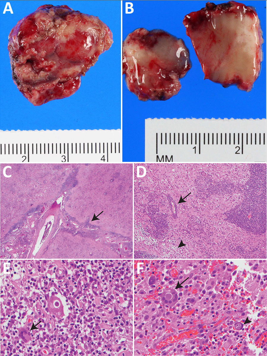
Figure 2. Resected occipital lesion gross and histopathology from patient in study of neurosurgical biopsy and resection for diagnosis and treatment of Balamuthia mandrillaris amebic encephalitis, United States. A) Gross pathology revealed multiple pink-tan, firm tissue fragments. B) Cut sections reveal a heterogenous, tan-pink, and soft cut surface with effacement of the white and gray matter demarcation. C, D) Hematoxylin and eosin stained sections of the brain show leptomeningeal thickening with striking perivascular lymphocytic infiltrate (C, arrow) extending into the cortical parenchyma with foci of lymphocytic infiltration into the vessel wall, consistent with vasculitis (D, arrow). The architecture of the cortical gray matter is obliterated by neuronal loss, reactive gemistocytic astrocytes and infiltration by macrophages, multinucleated giant cells, and lymphocytes (D, arrowhead). This involves the adjacent white matter with areas of parenchymal loss/liquefactive necrosis (D). Original magnification ×2 for panel C, ×10 for panel D. E, F) Many of the multinucleated giant cells (E, arrow) contain large round thick-walled parasites (F, arrow) containing granular material and an occasional inconspicuous, dot like nucleus (F, arrowhead). Original magnification ×40 for panels E and F. Occasional structures resembling engulfed organisms are seen in the parenchyma (D–F).

Main Article

1These authors contributed equally to this article.

Page created: June 10, 2026
Page updated: June 23, 2026
Page reviewed: June 23, 2026
The conclusions, findings, and opinions expressed by authors contributing to this journal do not necessarily reflect the official position of the U.S. Department of Health and Human Services, the Public Health Service, the Centers for Disease Control and Prevention, or the authors' affiliated institutions. Use of trade names is for identification only and does not imply endorsement by any of the groups named above.
file_external