Volume 32, Number 8—August 2026
Dispatch
Camel Prion Disease, Tataouine, Tunisia, 2019–2021

Figure 1
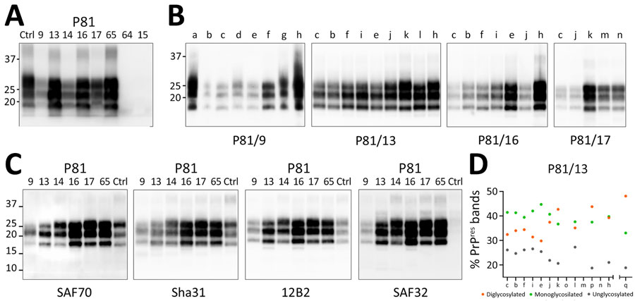
Figure 1. Detection and characterization of the proteinase K–resistant core (PrPres) of the pathological isoform of prion protein (PrPSc) from brain tissues of dromedary camels, Tunisia, 2019–2021. A) PrPres detected using TeSeE Western blot kit. Membrane probed with the Sha31 monoclonal antibody. Loading order (left to right): ovine scrapie control, Tunisian camel prion disease–positive cases (animal nos. P81/9–65), and Tunisian camel prion disease–negative samples (animal nos. P81/64 and P81/15). All samples, except for P81/64 and P81/15, were diluted 1:4 before electrophoresis and loaded at 3.75 mg tissue equivalent/lane. The negative samples were loaded at 15 mg tissue equivalent/lane. Approximate molecular weights (in kDa) are shown on the left side of the blot. B) Representative Western blot analysis of different available brain regions from selected positive cases. Brain tissues analyzed using the ISS discriminatory Western blot protocol. Case identifiers are indicated below each blot; corresponding brain regions are labeled at the top. All membranes were probed with the 12B2 antibody, except for P81/17, which was probed with L42. Tissue equivalents loaded per lane: 2 mg for P81/9 and 0.5 mg for P81/13, P81/16, and P81/17. Molecular weights (in kDa) are shown on the left side of the blot. C) Representative replica blots showing the epitope mapping analysis of PrPres from brain homogenates of positive dromedary camel cases. A preliminary assessment of dromedary PrPres reactivity with a panel of monoclonal antibodies was performed to identify suitable diagnostic tools for camel prion disease and to characterize PrPres. Within each antibody group, the antibody with the best sensitivity toward dromedary PrPSc was chosen for epitope mapping (Appendix Table 2, Figure 1). In addition, the SAF32 antibody, which targets the octarepeat region, was included to enable comparison with scrapie, in which that epitope is partially lost. Brain areas analyzed for each sample: P81/9, prefrontal cortex; P81/13, frontal cortex; P81/14, thalamus; P81/16, parietal cortex; P81/17, prefrontal cortex; P81/65, not identifiable; scrapie, medulla oblongata. A scrapie-affected sheep sample was included as control in last lane of each blot. Molecular weights (in kDa) are shown on the left side of the blot. Membranes were probed with monoclonal antibodies indicated below each blot. D) Relative proportion of diglycosylated, monoglycosylated, and unglycosylated PrPres bands in each available brain region of animal P81/13. Scrapie (q) is shown on the right for comparison. Quantifications were performed on membrane probed with 12B2 antibody. Brain regions: a, striatum; b, frontal cortex; c, prefrontal cortex; d, capsule; e, occipital cortex; f, parietal cortex; g, cerebellar peduncle; h, cerebellum; i, temporal cortex; j, basal ganglia; k, thalamus; l, hippocampus; m, midbrain; n, medulla oblongata; o, hypothalamus; p, pons. Ctrl, control.
1These authors contributed equally to this article.