Volume 32, Number 8—August 2026
Dispatch
Camel Prion Disease, Tataouine, Tunisia, 2019–2021

Figure 3
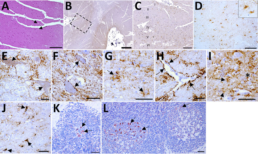
Figure 3. Histopathologic and immunohistochemical analyses of brain tissues and lymph nodes of camel prion disease–affected dromedary camels, Tunisia, 2019–2021. Examination of all available cerebral cortices (Appendix Table 3) showed mild spongiform changes exclusively in the temporal and occipital cortex (A) of sample P81/16. B) Immunostaining of the pathological isoform of prion protein (PrPSc) found in prefrontal cortex of sample P81/13. C) Magnification of the prefrontal cortex (dashed box in panel B) showing the involvement of the I and V–VI layers. D) Intraglial (inset) and intraneuronal PrPSc depositions observed in the thalamus in animal P81/9. E–I) PrPSc deposition patterns observed in camel prion disease–affected brain tissues: intraneuronal (E, arrows), perineuronal (F, arrows), glial associated (G, arrows), perivascular (H, arrows), punctate (I, arrows) and diffuse (I, asterisk) in the neuropil. J) Dense intra-astrocytic PrPSc deposition (arrows) observed in the medulla oblongata. K–L) PrPSc deposition, observed in primary and secondary follicles of lymph nodes, appeared as a reticular network within the center of lymphoid follicles, accompanied by fine to coarse cytoplasmic granules in nonlymphoid cells. Representative immunostaining (arrows) of mandibular lymph node from animal P81/65 (K) and prescapular lymph node from animal P81/17 (L) highlights intense granular PrPSc depositions in tingible body macrophages within germinal centers. Brown indicates PrPSc deposition in brain sections; red indicates PrPSc deposition in lymph nodes. Scale bars indicate 50 µm in panels A and D, 100 µm in panel B, 250 µm in panel C, and 20 µm in panels E–L.
1These authors contributed equally to this article.