Camel Prion Disease, Tataouine, Tunisia, 2019–2021
Abdelkader Amara
1, Michele Angelo Di Bari
1, Kéfia Elmehatli, Rosalia Bruno, Rihab Andolsi, Barbara Chiappini, Ilaria Vanni, Elena Esposito, Geraldina Riccardi, Obaid Allah Ben Abid, Stefano Marcon, Atef Malek, Boubaker Ben Smida, Haykel Kessa, Walid Chandoul, Mariem Handous, Roukaya Khorchani, Romolo Nonno, Malek Zrelli, Umberto Agrimi, Gabriele Vaccari, and Laura Pirisinu

Author affiliation: Université Mannouba Tunisie, École Nationale de Médecine Vétérinaire, Sidi Thabet, Tunisia (A. Amara, R. Andolsi, A. Malek); Istituto Superiore di Sanità, Rome, Italy (M.A. Di Bari, R. Bruno, B. Chiappini, I. Vanni, E. Esposito, G. Riccardi, O.A. Ben Abid, S. Marcon, R. Nonno, U. Agrimi, G. Vaccari, L. Pirisinu); Arrondissement de Production Animale de Siliana, Siliana, Tunisia (K. Emehatli); Regional Delegation for Agricultural Development in Tataouine, Tataouine, Tunisia (B. Ben Smida); Arrondissement de Production Animale de Sousse, Sousse, Tunisia (H. Kessa); Regional Delegation for Agriculture in Medenine, Medenine, Tunisia (W. Chandoul); Institut Pasteur de Tunisie, Tunis, Tunisia (M. Handous); Direction Générale des Services Vétérinaires, Ministère de l’Agriculture, Tunis (R. Khorchani) Ministry of Agriculture, Tunis (M. Zrelli)
Main Article
Figure 2
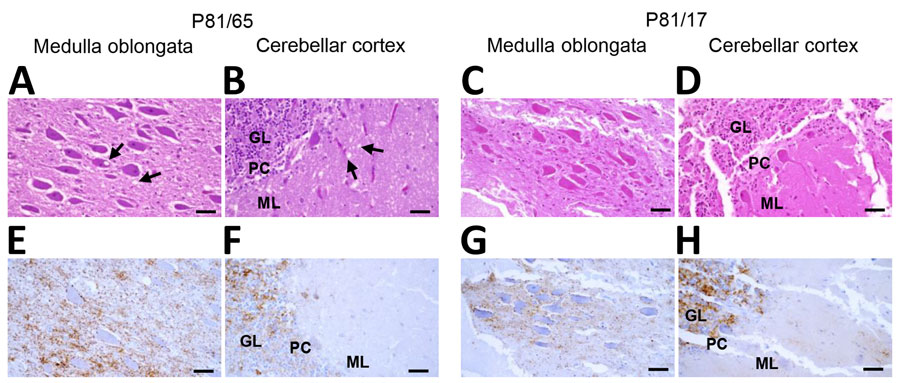
Figure 2. Histopathologic and immunohistochemical analyses of medulla oblongata and cerebellum of dromedary camels, Tunisia, 2019–2021. Images show the analyses performed on medulla oblongata (A, E, C, G) and cerebellum (B, F, D, H) of 2 representative animals (P81/65 and P81/17). Hematoxylin and eosin staining of the obex (A, C) and cerebellar cortex (B, D) revealed spongiform changes in the nucleus of the solitary tract (A, arrows) and in the molecular layer of cerebellar cortex (B, arrows) of P81/65, but no such alterations were observed in P81/17 (C, D). Conversely, immunohistochemistry revealed deposition of the pathological isoform of prion protein in both animals (E–H), affecting the nucleus of solitary tract (E, G) and cerebellar cortex (F and H). Brown color indicates PrPSc deposition. Brain sections from P81/17 showed partial loss of tissue integrity. Scale bars indicate 20 μm. GL, granular layer; ML, molecular layer; PC, Purkinje cells.
Main Article
Page created: May 21, 2026
Page updated: July 30, 2026
Page reviewed: July 30, 2026
The conclusions, findings, and opinions expressed by authors contributing to this journal do not necessarily reflect the official position of the U.S. Department of Health and Human Services, the Public Health Service, the Centers for Disease Control and Prevention, or the authors' affiliated institutions. Use of trade names is for identification only and does not imply endorsement by any of the groups named above.