Volume 32, Number 9—September 2026
Research Letter
Autochthonous Canine Pythiosis Linked with Freshwater Exposure, Italy, 2021–2024

 , Anna Rita Molinar Min, Patrizia Danesi, Francesco Albanese, Nicla Furiani, Moira Monaco, Fabiano Necci, Chiara Noli, Sofia Sgubin, Antonella Varina, and Mario Pasquetti
Cite This Article
Citation for Media
Abstract
We report 7 recognized autochthonous cases of canine pythiosis identified in Italy during 2021–2024. All infected dogs had freshwater exposure. Cases involved multiple lakes and occurred over consecutive years. Sequenced isolates clustered within clade IV of the Pythium insidiosum complex, currently designated Pythium periculosum.
Pythiosis is a severe disease caused by pathogenic oomycetes of the genus Pythium and is classically associated with tropical and subtropical climates and freshwater environments (1,2). The disease affects mainly humans, horses, and dogs and is typically linked to exposure to stagnant or slow-moving freshwater habitats that support production of infective zoospores. Most mammalian infections involve the Pythium insidiosum complex, although there have been occasional reports citing additional pathogenic oomycetes (3,4).
Increasing reports from regions outside traditionally recognized endemic areas, including northern areas of the United States and Japan, suggest a broader global distribution of pythiosis (2). Europe, however, remains exceptionally affected. A recent systematic review identified Spain as the only country in Europe in which locally acquired human pythiosis cases had been documented (2,5). In 2023, we reported 2 autochthonous canine cutaneous pythiosis cases in Italy, which were linked to recurrent exposure to Lake Bracciano in central Italy (6). Those initial observations suggested the possible presence of a local ecologic niche favorable to pathogenic Pythium species. We subsequently identified 5 additional canine cases diagnosed during 2021–2024, bringing the total number of recognized locally acquired canine cases in Italy to 7. None of the dogs had traveled outside Italy before disease onset, supporting the notion of locally acquired infection.
Figure 1
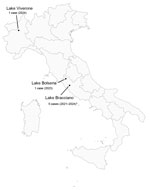
Figure 1. Freshwater sites associated with autochthonous canine pythiosis cases for a study of autochthonous canine pythiosis linked with freshwater exposure, Italy, 2021–2024. Dots indicate lakes linked to reported exposure histories. Labels...
All dogs had histories of repeated exposure to natural freshwater environments. Reported sites included Lake Bracciano (5 cases), Lake Bolsena (1 case), and Lake Viverone in northern Italy (1 case) (Figure 1). Distinct cases linked to Lake Bracciano were recognized from 2021 through 2024, suggesting persistent local environmental conditions favorable to pathogenic Pythium species. In addition, identification of a case associated with Lake Viverone expands the geographic range of recognized exposure sites by >300 km. Although environmental investigations were outside the scope of this study, all implicated sites feature shallow, near-shore areas that undergo marked seasonal warming during summer months. Such conditions may favor persistence or proliferation of pathogenic Pythium species (1). All factors considered, we believe that our findings support the presence of ecologic niches potentially suitable for pathogenic oomycetes in different regions of Italy despite the absence of traditionally recognized tropical climatic conditions.
Figure 2
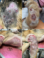
Figure 2. Representative cutaneous manifestations of canine pythiosis for a study of autochthonous canine pythiosis linked with freshwater exposure, Italy, 2021–2024. Lesions were characterized by chronic ulceration, nodules or plaques, draining tracts,...
Six of the 7 infected dogs were German shepherds. Although the number of cases remains limited, this predominance is noteworthy and suggest that factors beyond environmental exposure alone might contribute to disease occurrence. Published canine series from endemic regions show variable breed distributions, suggesting that disease emergence likely reflects a multifactorial interplay between environmental exposure, host-related factors, and clinical recognition patterns. Clinical manifestations were predominantly cutaneous and characterized by chronic nodules, plaques, ulceration, and draining tracts (Figure 2); 1 dog later developed gastrointestinal involvement. Researchers have described similar cutaneous and gastrointestinal manifestations in canine pythiosis occurring in other regions of the world (7–9). The predominance of cutaneous presentations in our series may reflect route of exposure through damaged skin or recognition bias, because gastrointestinal disease may be less readily diagnosed. In several cases, the initial clinical manifestation suggested inflammatory or bacterial disorders, and empirical treatment and, in some cases, repeated surgery preceded definitive diagnosis (Appendix).
We established diagnosis by combinations of cytology, histopathology, culture, and sequencing (Appendix). Cytologic and histologic examination showed broad, irregular, sparsely septate, hyphae-like structures associated with pyogranulomatous inflammation. Internal transcribed spacer sequence data were available for 6 cases, and those isolates clustered within clade IV of the P. insidiosum complex, corresponding to the lineage currently designated Pythium periculosum. (10) We conducted phylogenetic analysis using representative reference sequences spanning the 4 major clades recognized within the complex (Appendix Figure). All sequences from Italy grouped within clade IV, consistent with the broader extra-American representation previously observed for this clade (10). One additional case, for which only large subunit sequence data were available, showed highest similarity to isolates previously assigned to the same lineage. The sequences from Italy also showed limited divergence from one another and appeared closely related within the clade IV framework, suggesting a geographically related cluster.
Our findings provide further evidence that canine pythiosis can occur in temperate settings outside traditionally recognized endemic areas. Recurrent identification of cases associated with freshwater exposure suggests that suitable environmental niches may exist in multiple regions of Italy despite the absence of classically tropical climatic conditions. From a One Health perspective, dogs may act as sentinels of environmental exposure risk relevant to multiple hosts and may contribute to recognition of previously underappreciated ecologic niches favorable to pathogenic oomycetes. Increased awareness among veterinary and human healthcare professionals will be important for earlier recognition of this emerging disease in Europe.
Dr. Peano is an assistant professor in the Department of Veterinary Sciences, University of Turin, Italy. His research focuses on fungal diseases of domestic and wild animals, with particular interest in their epidemiology and One Health implications.
Acknowledgment
The authors used ChatGPT (OpenAI, https://www.openai.com) for language editing and technical guidance in figure formatting but performed all data collection, analyses, interpretations, and figure preparation themselves. No images, data, or scientific content were generated by AI.
References
- Gaastra W, Lipman LJ, De Cock AW, Exel TK, Pegge RB, Scheurwater J, et al. Pythium insidiosum: an overview. Vet Microbiol. 2010;146:1–16. DOIPubMedGoogle Scholar
- Yolanda H, Krajaejun T. Global distribution and clinical features of pythiosis in humans and animals. J Fungi (Basel). 2022;8:182. DOIPubMedGoogle Scholar
- Calvano TP, Blatz PJ, Vento TJ, Wickes BL, Sutton DA, Thompson EH, et al. Pythium aphanidermatum infection following combat trauma. J Clin Microbiol. 2011;49:3710–3. DOIPubMedGoogle Scholar
- Farmer AR, Murray CK, Driscoll IR, Wickes BL, Wiederhold N, Sutton DA, et al. Combat-related Pythium aphanidermatum invasive wound infection: case report and discussion of utility of molecular diagnostics. J Clin Microbiol. 2015;53:1968–75. DOIPubMedGoogle Scholar
- Bernheim D, Dupont D, Aptel F, Dard C, Chiquet C, Normand AC, et al. Pythiosis: case report leading to new features in clinical and diagnostic management of this fungal-like infection. Int J Infect Dis. 2019;86:40–3. DOIPubMedGoogle Scholar
- Peano A, Min ARM, Fondati A, Romano E, Brachelente C, Porcellato I, et al. Cutaneous pythiosis in 2 dogs, Italy. Emerg Infect Dis. 2023;29:1447–50. DOIPubMedGoogle Scholar
- Oldenhoff W, Grooters A, Pinkerton ME, Knorr J, Trepanier L. Cutaneous pythiosis in two dogs from Wisconsin, USA. Vet Dermatol. 2014;25:52–e21. DOIPubMedGoogle Scholar
- Berryessa NA, Marks SL, Pesavento PA, Krasnansky T, Yoshimoto SK, Johnson EG, et al. Gastrointestinal pythiosis in 10 dogs from California. J Vet Intern Med. 2008;22:1065–9. DOIPubMedGoogle Scholar
- Billings P, Walton S, Shmalberg J, Santoro D. The use of mefenoxam to treat cutaneous and gastrointestinal pythiosis in dogs: a retrospective study. Microorganisms. 2023;11:1726. DOIPubMedGoogle Scholar
- Miraglia BM, Mendoza L, Rammohan R, Vilela L, Vilela C, Vilela G, et al. Pythium insidiosum complex hides a cryptic novel species: Pythium periculosum. Fungal Biol. 2022;126:366–74. DOIPubMedGoogle Scholar
Figures
Cite This ArticleOriginal Publication Date: August 17, 2026
Table of Contents – Volume 32, Number 9—September 2026
| EID Search Options |
|---|
 Advanced Article Search – Search articles by author and/or keyword.
|
 Articles by Country Search – Search articles by the topic country.
|
 Article Type Search – Search articles by article type and issue.
|
Please use the form below to submit correspondence to the authors or contact them at the following address:
Andrea Peano, Dipartimento di Scienze Veterinarie, Largo P. Braccini 2, 10095 Grugliasco, Torino, Italy
Top