Volume 32, Number 9—September 2026
Research
Reemergence of Scrub Typhus Associated with Leptotrombidium akamushi Mites and Karp-Like Orientia tsutsugamushi Genotype Bacteria, Japan, 2024–2025
 , Nobuhiro Takada, Takashi Katayama, Yuki Hashimoto, Kei Nagano, Hirotaka Komine, and Yukihiro Akeda
Cite This Article
Citation for Media
Abstract
Scrub typhus is a miteborne infectious disease caused by Orientia tsutsugamushi bacteria. In Japan, most cases occur in spring and autumn and are transmitted by Leptotrombidium pallidum and L. scutellare mites; summer scrub typhus, transmitted by L. akamushi mites, is rarely reported. In August 2024, a pregnant woman had scrub typhus develop after attending a fireworks event in Niigata Prefecture, Japan, resulting in fetal death. A Vietnam-related Karp-like O. tsutsugamushi genotype, distinct from the Kato type usually associated with L. akamushi mites, was detected from patient samples. Field investigations at the site of the fireworks event revealed seasonal predominance of L. akamushi mites in summer. We detected O. tsutsugamushi sequences identical to the patient’s strain in 3 L. akamushi mite larvae and 1 field mouse, suggesting that L. akamushi mites were the source of human infection. Our findings suggest reemergence of summer scrub typhus associated with infected L. akamushi mites carrying a locally novel genotype.
Scrub typhus is a miteborne infectious disease caused by Orientia tsutsugamushi bacteria and transmitted by larval trombiculid mites of the genus Leptotrombidium (1). Patients typically have an eschar develop at the site of the mite bite, accompanied by fever, rash, and elevated liver enzyme and C-reactive protein levels (2,3). Tetracycline-class antimicrobial agents are generally highly effective against scrub typhus, but delayed treatment can lead to severe disease and occasionally death.
In Japan, scrub typhus exhibits a bimodal seasonal distribution, with peaks occurring in spring and autumn (2). That seasonal pattern is largely determined by the life cycles of vector mite species. The principal vectors in Japan are L. pallidum and L. scutellare mites (1,4). Both species hatch in autumn, and their larvae parasitize vertebrate hosts, contributing to the autumn peak of disease incidence. L. pallidum larvae can overwinter and remain active until the following spring, thereby contributing to the spring peak of scrub typhus cases. Historically, summer scrub typhus, transmitted by L. akamushi mites, occurred in river basin environments of northern and central Japan, including major river systems in Akita, Yamagata, and Niigata Prefectures, such as the Shinano River Basin (2,5). However, summer scrub typhus is now rare, and only sporadic cases have been reported in recent decades, including a suspected Kato-type infection reported in Akita Prefecture in 2008 (6).
Different Leptotrombidium mite species are associated with specific O. tsutsugamushi serotypes. For example, L. akamushi mites are primarily associated with the Kato serotype, whereas L. pallidum and L. scutellare mites are associated with other major serotypes, including Gilliam, Karp, Kawasaki, and Kuroki (2,7,8). More recently, L. palpale mites have been proposed as a vector candidate for the Shimokoshi type O. tsutsugamushi bacteria (9). In Japan, reports of infections caused by the Kato serotype are rare, and the O. tsutsugamushi bacterial genotypes currently associated with L. akamushi mites remain poorly characterized in regions where summer scrub typhus was historically endemic and where that mite species was the principal vector.
In August 2024, a pregnant woman who had attended a fireworks event on a riverbank in Niigata Prefecture had scrub typhus develop, resulting in fetal death. She had no recent travel history outside Niigata Prefecture before symptom onset. O. tsutsugamushi bacteria were detected in the patient’s serum and placenta. Sequence analysis of the 56-kDa type–specific antigen (TSA) gene revealed a Karp-like genotype closely related to strains previously reported from Vietnam (10). The fireworks event, the suspected exposure site, was held in the Shinano River Basin, a region historically known for summer scrub typhus transmitted by L. akamushi mites.
Detection of O. tsutsugamushi in larval mites can provide evidence of the direct source of human infection, and infection in rodents can provide indirect evidence of infected mites in the area. Therefore, we conducted a field investigation in the suspected exposure area to capture mites and field mice to determine O. tsutsugamushi infection and characterize the local maintenance of O. tsutsugamushi in mite vectors and their host species.
Figure 1

Figure 1. Study sites for investigation of reemergence of scrub typhus associated with Leptotrombidium akamushi mites and Karp-like Orientia tsutsugamushigenotype bacteria, Japan, 2024–2025. Map shows suspected exposure site...
We conducted a field investigation at the suspected exposure site along a riverbank in Nagaoka City, Niigata Prefecture, Japan (37°27.283′N, 138°50.269′E). We selected 2 sampling sites within ≈200 meters of the suspected exposure site: site A was an area of dense bushy vegetation adjacent to farmland, and site B was an area of dense bushy vegetation immediately adjacent to the river’s edge (Figure 1). Both sites provided suitable habitats for trombiculid mites and small mammal hosts, including Apodemus spp. field mice.
We conducted field surveys at 1 or both sites during June, July, August, and November 2025; site A was not sampled in August. We used Sherman traps to capture field mice and set ≈50 traps per night across both sites. We anesthetized captured field mice and collected blood by cardiac puncture for serologic testing before humanely euthanizing the mice. We then collected spleen samples for molecular detection. We carefully removed attached larval trombiculid mites from field mice for mite species identification and O. tsutsugamushi detection. The Institutional Animal Care and Use Committee of the National Institute of Infectious Diseases approved all animal procedures (approval no. 125159).
Mite Identification
We morphologically identified mites under a stereomicroscope, according to standard taxonomic methods (11). Because L. akamushi mites have rarely been reported in recent field surveys, we performed molecular identification on selected specimens on the basis of the cytochrome oxidase I (COX1) gene (12) to verify species identification. We used PrepMan Ultra Sample Preparation Reagent (Thermo Fisher Scientific, https://www.thermofisher.com) to extract DNA from individual mites.
Detection of O. tsutsugamushi in Mice and Mites
We tested mouse serum samples for 6 O. tsutsugamushi antibody serotypes by using an indirect immunofluorescence assay (13) and considered titers >80 positive. We extracted DNA from spleen samples by using a FavorPrep Tissue Genomic DNA Extraction Mini Kit (Favorgen Biotech Corp., https://www.favorgen.com). We used real-time PCR targeting the 16S rRNA gene (14) on DNA extracted from field mouse spleens and engorged mites to screen for O. tsutsugamushi. For samples that tested positive or equivocal, we conducted further analysis by using nested PCR targeting the 56-kDa TSA gene (15). We sequenced amplicons and performed phylogenetic analysis by using the neighbor-joining method in MEGA X version 11.01.3 (https://www.megasoftware.net) with 1,000 bootstrap replicates (16).
Detection of O. tsutsugamushi in Mice
Figure 2
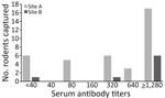
Figure 2. Antibody titers from field mice in a study of reemergence of scrub typhus associated with Leptotrombidium akamushi mites and Karp-like Orientia tsutsugamushigenotype bacteria, Japan, 2024–2025. We...
We captured a total of 54 field mice, all of which were large Japanese field mice (Apodemus speciosus). Serum samples suitable for serologic testing were obtained from 45 mice (37 from site A and 8 from site B). From site A, 31 (84%) of 37 tested field mice were O. tsutsugamushi–seropositive. In site B, 7 (88%) of 8 tested field mice were O. tsutsugamushi–seropositive (Figure 2). From site A, PCR genotyping revealed Japanese Gilliam–type strains of O. tsutsugamushi bacteria in field mice collected during June, July, and November, and we detected a Karp-type strain in 1 field mouse collected in June. At site B, we detected Japanese Gilliam–type strains in 1 field mouse in June.
Mite Abundance and Identification
Figure 3
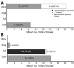
Figure 3. Seasonal abundance and species composition of trombiculid mites collected from field mice in a study of reemergence of scrub typhus associated with Leptotrombidium akamushi mites and Karp-like ...
The composition and seasonal abundance of mites collected from field mice differed markedly between the 2 sampling sites. From site A, we detected mites in November (mean 16.3 mites/mouse) and June (mean 6.5 mites/mouse) but not in July (Table). We conducted no sampling at site A in August. All mites collected in June were L. pallidum, whereas 57.7% of mites collected in November were L. pallidum and 42.3% were Neotrombicula japonica (Figure 3). At site B, all mites collected in June and August were L. pallidum, whereas L. akamushi accounted for 96.3% of mites collected in July (Figure 3).
Figure 4
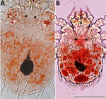
Figure 4. Comparison of engorged larval mites from a study of reemergence of scrub typhus associated with Leptotrombidium akamushi mites and Karp-like Orientia tsutsugamushi genotype bacteria, Japan, 2024–2025. A) ...
Representative mite specimens showed the characteristic position of the sensillary setae anterior to the posterolateral setae and 8 dorsal idiosomal setae, consistent with L. akamushi mites (Figure 4). Of L. akamushi 52 mites analyzed, 24 yielded COX1 sequences confirming species identification. The other 28 specimens did not yield reliable sequences, likely because of the extremely small body size of larvae and variable DNA quality. However, all specimens exhibited morphologic characteristics consistent with established diagnostic criteria for L. akamushi mites under stereomicroscopic examination. Together with the molecular results, those findings indicate the presence of L. akamushi mites at the study site.
Phylogenetic Analysis of O. tsutsugamushi Strains
We identified Japanese Gilliam–type O. tsutsugamushi strains in 5 field mice collected at site A in June, 1 field mouse collected in July, and 2 field mice collected in November. At site B, we identified the same strain in 1 field mouse and 1 mite collected in June. We identified a Karp-like strain of O. tsutsugamushi in 1 field mouse and 3 L. akamushi larvae collected at site B in July. COX1 analysis confirmed the larvae as L. akamushi species mites.
Figure 5
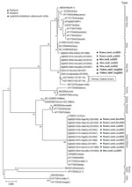
Figure 5. Phylogenetic relationships of Orientia tsutsugamushi strains detected in field mice, mites, and a human patient from a study of reemergence of scrub typhus associated with Leptotrombidium akamushi...
The Karp-like strain of O. tsutsugamushi we detected was more closely related to strains from Vietnam than to the Kato type historically associated with L. akamushi mites in Japan. The longest 56-kDa TSA gene sequence (959 bp) we obtained was from the placenta specimen (GenBank accession no. LC912005). That sequence showed the highest nucleotide identity (98.5%) to 2 previously reported strains from Vietnam (GenBank accession nos. HQ718457.1 [945 bp] and HQ718455.1 [959 bp]) (Figure 5). Related Karp-like sequences have been identified elsewhere in Asia, but those 2 strains from Vietnam had the highest sequence similarity to sequence LC912005. In addition, the 959-bp sequence obtained from the placenta specimen was identical to 2 other sequences (GenBank accession nos. LC912009 and LC912010) obtained from 2 L. akamushi mite larvae. The nucleotide sequences of an ≈420-bp region were identical among the placenta specimen (GenBank accession no. LC912005), patient serum sample (accession no. LC921007), 1 field mouse spleen sample (accession no. LC912003), and 3 L. akamushi larvae (accession nos. LC912007, LC912009, and LC912010). Phylogenetic analysis on the basis of that 420-bp region placed all samples within the same Karp-like cluster (Figure 5).
The O. tsutsugamushi–positive larvae were engorged L. akamushi larvae attached to field mice at the time of collection. However, the spleens of the field mouse hosts to which those larvae were attached tested negative for O. tsutsugamushi by PCR. Because trombiculid larvae feed only once on vertebrate hosts, larvae that acquire O. tsutsugamushi from an infected host cannot subsequently transmit the pathogen to another host. Moreover, horizontally acquired infection is not efficiently maintained across generations (8,17). Therefore, detection of O. tsutsugamushi in engorged larvae attached to PCR-negative field mice strongly suggests that those larvae acquired the pathogen before attaching to the mice, likely through vertical transmission; therefore, L. akamushi could be the source of human infection in this instance. Furthermore, sequence analysis showed that the nucleotide sequences of the O. tsutsugamushi bacterial strain in the larvae were identical to those detected in the patient’s serum and placenta, providing strong evidence that human infection was likely caused by bites of infected L. akamushi larvae during the fireworks event on the riverbank.
Our findings demonstrate marked seasonal differences in mite species abundance and composition within a limited area along a riverbank in Niigata Prefecture and identified L. akamushi as the predominant mite species at the time and location where the patient is suspected to have been exposed. Detection of the same genotype of O. tsutsugamushi in L. akamushi mites, field mice, and the patient suggests that human infection occurred at this site.
The marked predominance of L. akamushi in July suggests that this species is strongly associated with the summer occurrence of O. tsutsugamushi scrub typhus at this site. Historically, L. akamushi mites have been recognized as the principal vector of summer scrub typhus in northern Japan, particularly in river basin environments. The contrasting predominance of L. pallidum mites at site A and L. akamushi mites at site B might reflect differences in local environmental conditions, although the specific factors responsible remain unclear.
Of note, we identified engorged O. tsutsugamushi–positive larvae attached to field mice whose spleens tested O. tsutsugamushi–negative by PCR, indicating that the larvae acquired the pathogen before attaching to their hosts. Because trombiculid larvae feed only once on vertebrate hosts and horizontally acquired infection is not efficiently maintained across generations, only larvae that already harbor O. tsutsugamushi, presumably through vertical transmission, can serve as the source of human infection. Those findings suggest that O. tsutsugamushi is maintained primarily within mite populations and that infected larval mites are the principal source of human infection, rather than vertebrate hosts serving as amplification reservoirs.
The strain we identified was a Vietnam-related Karp-like genotype, distinct from the Kato type historically associated with L. akamushi mites, suggesting the presence of a previously unrecognized lineage associated with this vector species in Japan. That finding demonstrates that L. akamushi mites are not associated exclusively with the Kato genotype, as previously thought, but can also harbor genetically distinct lineages, including Vietnam-related Karp-like strains.
In summary, the high seroprevalence in field mice at both survey sites suggests widespread circulation of O. tsutsugamushi and infected vector mites within the study area. Collectively, our findings provide evidence for the re-emergence of summer scrub typhus associated with infected L. akamushi mites and highlight the need for continued vector surveillance and molecular O. tsutsugamushi bacterial surveillance to clarify scrub typhus risk in Japan.
Dr. Ogawa is a senior researcher in the Department of Bacteriology I at the National Institute of Infectious Diseases, Tokyo, Japan. His research interests include vector-borne infectious diseases and the molecular epidemiology of rickettsial pathogens.
Acknowledgment
We thank the Niigata Prefectural Government for granting permission to conduct rodent trapping at the study sites and colleagues at the National Institute of Infectious Diseases for support during field investigations. We are grateful to T. Miura for assistance with laboratory testing, patient information, and coordination with the attending physician. We thank Editage (https://www.editage.jp) for English language editing.
References
- Elliott I, Pearson I, Dahal P, Thomas NV, Roberts T, Newton PN. Scrub typhus ecology: a systematic review of Orientia in vectors and hosts. Parasit Vectors. 2019;12:513. DOIPubMedGoogle Scholar
- Kawamura A, Tanaka H, Tamura A, editors. Tsutsugamushi disease. Tokyo: University of Tokyo Press; 1995.
- Ogawa M, Hagiwara T, Kishimoto T, Shiga S, Yoshida Y, Furuya Y, et al. Scrub typhus in Japan: epidemiology and clinical features of cases reported in 1998. Am J Trop Med Hyg. 2002;67:162–5. DOIPubMedGoogle Scholar
- Iwasa M, Kasuya S, Noda N, Hioki A, Ito A, Ohtomo H. Trombiculid mites (Acari: Trombiculidae) and Rickettsia tsutsugamushi isolated from wild rodents in a new endemic area of Japan. J Med Entomol. 1990;27:501–8. DOIPubMedGoogle Scholar
- Kitaoka M, Asanuma K, Otsuji J. Transmission of Rickettsia orientalis to man by Leptotrombidium akamushi at a scrub typhus endemic area in Akita Prefecture, Japan. Am J Trop Med Hyg. 1974;23:993–9. DOIPubMedGoogle Scholar
- Sato H, Kokusho Y, Shibata C, Saito H, Saito S, Fujita H, et al. A case of classical tsutsugamushi disease confirmed after an interval of 15 years in Akita Prefecture, Japan [in Japanese]. Kansenshogaku Zasshi. 2010;84:454–6. DOIPubMedGoogle Scholar
- Takahashi M, Misumi H, Urakami H, Nakajima S, Furui S, Yamamoto S, et al. Mite vectors (Acari: Trombiculidae) of scrub typhus in a new endemic area in northern Kyoto, Japan. J Med Entomol. 2004;41:107–14. DOIPubMedGoogle Scholar
- Takahashi M, Murata M, Misumi H, Hori E, Kawamura A Jr, Tanaka H. Failed vertical transmission of Rickettsia tsutsugamushi (Rickettsiales: Rickettsiaceae) acquired from rickettsemic mice by Leptotrombidium pallidum (Acari: trombiculidae). J Med Entomol. 1994;31:212–6. DOIPubMedGoogle Scholar
- Seto J, Suzuki Y, Otani K, Qiu Y, Nakao R, Sugimoto C, et al. Proposed vector candidate: Leptotrombidium palpale for Shimokoshi type Orientia tsutsugamushi. Microbiol Immunol. 2013;57:111–7. DOIPubMedGoogle Scholar
- Duong V, Mai TT, Blasdell K, Lo V, Morvan C, Lay S, et al. Molecular epidemiology of Orientia tsutsugamushi in Cambodia and Central Vietnam reveals a broad region-wide genetic diversity. Infect Genet Evol. 2013;15:35–42. DOIPubMedGoogle Scholar
- Takada N, Takahashi M, Fujita H, Natsuaki M. Medical acarology in Japan [in Japanese]. Tokyo: Hokuryukan; 2019.
- Ogawa M, Takahashi M, Matsutani M, Takada N, Noda S, Saijo M. Obligate intracellular bacteria diversity in unfed Leptotrombidium scutellare larvae highlights novel bacterial endosymbionts of mites. Microbiol Immunol. 2020;64:1–9. DOIPubMedGoogle Scholar
- Ogawa M, Ando S, Saijo M. Evaluation of recombinant type-specific antigens of Orientia tsutsugamushi expressed by a baculovirus-insect cell system as antigens for indirect immunofluorescence assay in the serological diagnosis of scrub typhus. Jpn J Infect Dis. 2020;73:330–5. DOIPubMedGoogle Scholar
- Kawamori F, Shimazu Y, Sato H, Monma N, Ikegaya A, Yamamoto S, et al. Evaluation of diagnostic assay for rickettsioses using duplex real-time PCR in multiple laboratories in Japan. Jpn J Infect Dis. 2018;71:267–73. DOIPubMedGoogle Scholar
- Furuya Y, Yoshida Y, Katayama T, Kawamori F, Yamamoto S, Ohashi N, et al. Specific amplification of Rickettsia tsutsugamushi DNA from clinical specimens by polymerase chain reaction. J Clin Microbiol. 1991;29:2628–30. DOIPubMedGoogle Scholar
- Kumar S, Stecher G, Li M, Knyaz C, Tamura K. MEGA X: molecular evolutionary genetics analysis across computing platforms. Mol Biol Evol. 2018;35:1547–9. DOIPubMedGoogle Scholar
- Takahashi M, Murata M, Nogami S, Hori E, Kawamura A Jr, Tanaka H. Transovarial transmission of Rickettsia tsutsugamushi in Leptotrombidium pallidum successively reared in the laboratory. Jpn J Exp Med. 1988;58:213–8.PubMedGoogle Scholar
Figures
Table
Cite This ArticleOriginal Publication Date: August 18, 2026
Table of Contents – Volume 32, Number 9—September 2026
| EID Search Options |
|---|
 Advanced Article Search – Search articles by author and/or keyword.
|
 Articles by Country Search – Search articles by the topic country.
|
 Article Type Search – Search articles by article type and issue.
|
Please use the form below to submit correspondence to the authors or contact them at the following address:
Motohiko Ogawa, Department of Bacteriology I, National Institute of Infectious Diseases, Japan Institute for Health Security, 1-23-1, Toyama, Shinjuku-ku, Tokyo 162-8640, Japan
Top