Volume 13, Number 6—June 2007
Research
Levels of Abnormal Prion Protein in Deer and Elk with Chronic Wasting Disease
Figure 2
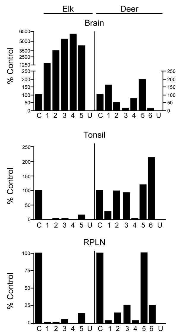
Figure 2. Quantification of disease-associated prion protein (PrPres) in brain, tonsil, and retropharyngeal lymph node (RPLN) from chronic wasting disease–affected elk and deer. The relative amount of PrPres in each lane of Figure 1 is shown relative to a common control described in the Figure 1 legend. A split scale is shown for elk brain because the PrPres signal from each elk brain was strong enough with 2-mg equivalents of tissue to obscure the protein patterns on the gel. Twenty-milligram equivalents were analyzed for all other tissues. The 10-fold difference in the amount of tissue equivalents loaded is accounted for by the split scale, where the result from 2-mg equivalents was multiplied by 10. Data shown are the average of 4 duplicate gels run for each sample. PrPres level in elk brain is significantly different from deer brain (p<0.001), elk tonsil is significantly different from deer tonsil (p = 0.0274), and elk RPLN is significantly different from deer RPLN (p = 0.0087) (Mann-Whitney test). C, reference control; U, uninfected elk or deer.