Volume 15, Number 1—January 2009
Dispatch
Hepatitis E Virus Genotype 3 Diversity, France
Abstract
We characterized 42 hepatitis E virus (HEV) genotype 3 strains from infected patients in France in 3 parts of the genome and sequenced the full-length HEV genotype 3f genome found in Europe. These strains are closely related to swine strains in Europe, which suggests zoonotic transmission of HEV in France.
Hepatitis E is a water-borne infection in developing countries and is believed to spread zoonotically in industrialized countries (1). Hepatitis E virus (HEV) is a positive-sense RNA virus that belongs to the family Hepeviridae (2). The coding region consists of 3 discontinuous open reading frames (ORFs). One region within ORF1, the hypervariable region, displays substantial genetic diversity (2). HEV strains are classified into 4 genotypes, and it was recently proposed that HEV genotypes are divided into 24 subtypes (3). Although genotypes 1 and 2 have been found exclusively in humans, genotypes 3 and 4 have been found in humans and animals such as pigs, boars, and deer. Genotypes 1 and 2 have been isolated in tropical and subtropical countries in Asia, Africa, and America; genotype 4 has been found only in Asia. Genotype 3 has been identified almost worldwide, but the distribution of its 10 subtypes varies greatly. Subtypes 3a and 3j strains have been mainly identified in North America; 3b, 3d, and 3g strains in Asia; and 3c, 3e, 3f, 3h, and 3i strains in Europe (3). The finding of several cases of HEV infection in southern France (4,5) prompted us to characterize the diversity of the strains and to determine the full-length sequence of the most prevalent genotype in this area.
We studied 42 HEV strains from patients at Toulouse University Hospital, France. None of the patients had traveled abroad during the previous 6 months or reported any contact with pigs before the onset of the disease. HEV infection was determined by detecting HEV RNA by using molecular tools (5). We sequenced 2 fragments within ORF1, the hypervariable region, and the RNA-dependent RNA polymerase (RdRp) genes as previously described (6,7). A 348-nt fragment of ORF2 was amplified according to the protocol of Inoue et al. (8). The whole genome was amplified with 6 overlapping reverse transcription–PCRs. The primers are listed in Table 1. The reverse transcription conditions were 50°C for 30 min and 85°C for 5 min. The PCR cycling conditions were initial denaturation at 94°C for 2 min, then 50 cycles of denaturation at 94°C for 15 sec, annealing at 60°C for 30 sec, and elongation at 68°C for 3 min.
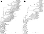
The phylogenetic tree obtained for the ORF2 region showed that all strains belonged to genotype 3 (Figure 1, panel A). A total of 37 strains (TLS1–TLS37) segregated as a distinct clade among genotype-3 reference sequences, whereas 5 other strains (TLS38–TLS42) formed distinct branches in the tree. TLS1–TLS37 could be subtyped as 3f, according to the classification proposed by Lu et al. (3). The nucleotide sequences of our local 3f strains and the strains from Spain and the Netherlands, which had been identified in humans, pigs, and sewage of animal origin, had 90.2%–95.3% identity. The 5 remaining strains were also genetically related to swine strains. Strains TLS38 and TLS42 were more closely related to 3e strains from swine in Great Britain (88.5%–92.1% nucleotide sequence identity). These strains were also closely related to Japanese strains AB248520 and AB248522, which may have a British origin (87.5%–93.4% identity) (9). Strains TLS40 and TLS41 were located on the same branch as swine HEV isolates identified in the Netherlands and classified as subtype 3c. The nucleotide sequence of TLS41 had 91.7%–92.7% identity with that of strains from pigs in the Netherlands, whereas that of TLS40 was only 85.5%–86.1% identical to that of pig strains from the Netherlands. Strain TLS39 was similar to the 3b strains identified in Japan in human patients or animals (86.8%–90.1% nucleotide sequence identity).
The topology of the phylogenetic tree obtained for the RdRp region was similar to that of ORF2 (Figure 1, panel B). All the strains clustered with genotype 3 strains. The same 37 strains (TLS1–TLS37) clustered together (87.9%–99.1% nucleotide sequence identity) and the same 5 strains (TLS38–TLS42) appeared to be more divergent.
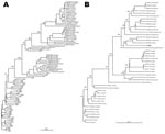
The hypervariable region gave the same clade of 37 strains in the phylogenetic tree, whereas the 5 remaining strains were more divergent (Figure 2, panel A). Strains TLS1–TLS37 exhibited only 70.1%–86.8% nucleotide sequence identity in this part of the genome, which shows the great diversity of this region. According to the primers position (6), a fragment of 345 nt was expected for all the strains. Actually, the fragments obtained after amplification of the hypervariable region of the genotype 3f strains varied from 435 nt for 29 samples, to 412 nt for 3 samples, and to 348 nt for 5 samples. For the 3e strains, the fragments were 387 nt and 412 nt. The PCR product from the hypervariable region was 345 nt for only the 3c and 3b strains.
The complete genome from TLS25 strain (genotype 3f) was amplified. The genomic length was 7,321 nt with the 3 major ORFs. Comparison with full-length reference sequences showed an insertion of ≈90 nt in the hypervariable region. Phylogenetic analysis of the sequences of HEV genotypes 1–4 strains confirmed that TLS25 belonged to genotype 3 and was genetically distinct from the genotype 3 strains found in Asia and in the United States (Figure 2, panel B). Comparisons with the complete genome sequences of HEV indicated that strain TLS25 shared 72.0%–72.9% nucleotide sequence identity with genotype 1 strains, 72% nucleotide sequence identity with genotype 2 strains, and 73.7%–74.5% nucleotide sequence identity with genotype 4 strains (Table 2). Strain TLS25 shows 83.1%–84.3% nucleotide sequence identity with HEV genotype 3e, 81.8% identity with the HEV strain 3g, 79.7%–80.3% identity with HEV genotype 3b, and 79.5%–81.8% identity with HEV genotype 3a over the entire genome. No recombination event was detected within the TLS25 strain by using the Recombinant Detection Program (http://darwin.uvigo.es/rdp/rdp.html). The sequences were deposited in GenBank under accession nos. EU495106–EU495232.
Most (88%) of the HEV strains in France belonged to 3f subtype, but 3c, 3e, and 3b strains were also identified. Phylogenetic analyses indicated that HEV strains in France were related to swine strains previously identified in Europe. This full-length sequencing of an HEV genotype 3 strain from Europe showed it to be distinct from the genotype 3 strains found on other continents, which illustrates the great diversity of this genotype. Due to an insertion in the hypervariable region, the TLS25 sequence is longer than other HEV sequences available in GenBank. A variation in the length of the hypervariable region in 1 human strain of genotype 3e was previously reported (9). Because the function of this region of the genome is still unknown, the effect of such insertions on virus biology has yet to be elucidated. Zhai et al. reported that phylogenetic analyses within the RdRp region correlated well with the results from the phylogenetic analyses of the complete genome (7), whereas Lu et al. found that the ORF2 region was the region that determined most accurately genotypes and subtypes (3). Our data indicate that both regions can be used to determine the genotype and subtype. The source of autochthonous hepatitis E infection in industrialized countries is unknown. One hypothesis, supported by molecular epidemiologic studies, is that it is an emerging zoonotic infection (10,11). Contamination with HEV may be linked to occupational exposure (12,13), consumption of undercooked meat (14,15), or exposure to a contaminated environment.
Our study characterized several human HEV strains and sequenced the full-length HEV 3f genome. These strains were closely related to European swine strains. Prospective in-depth epidemiologic studies based on structured interviews are ongoing to clarify the routes of transmission in southwest France.
Dr Legrand-Abravanel is a researcher in the Virology Department at Tououse University Hospital. Her primary research interest is the genetic variability of hepatitis viruses.
References
- Okamoto H. Genetic variability and evolution of hepatitis E virus. Virus Res. 2007;127:216–28. DOIPubMedGoogle Scholar
- Lu L, Li C, Hagedorn CH. Phylogenetic analysis of global hepatitis E virus sequences: genetic diversity, subtypes and zoonosis. Rev Med Virol. 2006;16:5–36. DOIPubMedGoogle Scholar
- Kamar N, Selves J, Mansuy JM, Ouezzani L, Peron JM, Guitard J, Hepatitis E virus and chronic hepatitis in organ-transplant recipients. N Engl J Med. 2008;358:811–7. DOIPubMedGoogle Scholar
- Mansuy JM, Peron JM, Abravanel F, Poirson H, Dubois M, Miedouge M, Hepatitis E in the south west of France in individuals who have never visited an endemic area. J Med Virol. 2004;74:419–24. DOIPubMedGoogle Scholar
- Kabrane-Lazizi Y, Zhang M, Purcell RH, Miller KD, Davey RT, Emerson SU. Acute hepatitis caused by a novel strain of hepatitis E virus most closely related to United States strains. J Gen Virol. 2001;82:1687–93.PubMedGoogle Scholar
- Zhai L, Dai X, Meng J. Hepatitis E virus genotyping based on full-length genome and partial genomic regions. Virus Res. 2006;120:57–69. DOIPubMedGoogle Scholar
- Inoue J, Takahashi M, Yazaki Y, Tsuda F, Okamoto H. Development and validation of an improved RT-PCR assay with nested universal primers for detection of hepatitis E virus strains with significant sequence divergence. J Virol Methods. 2006;137:325–33. DOIPubMedGoogle Scholar
- Inoue J, Takahashi M, Ito K, Shimosegawa T, Okamoto H. Analysis of human and swine hepatitis E virus (HEV) isolates of genotype 3 in Japan that are only 81–83% similar to reported HEV isolates of the same genotype over the entire genome. J Gen Virol. 2006;87:2363–9. DOIPubMedGoogle Scholar
- Meng XJ, Purcell RH, Halbur PG, Lehman JR, Webb DM, Tsareva TS, A novel virus in swine is closely related to the human hepatitis E virus. Proc Natl Acad Sci U S A. 1997;94:9860–5. DOIPubMedGoogle Scholar
- Banks M, Bendall R, Grierson S, Heath G, Mitchell J, Dalton H. Human and porcine hepatitis E virus strains, United Kingdom. Emerg Infect Dis. 2004;10:953–5.PubMedGoogle Scholar
- Colson P, Kaba M, Bernit E, Motte A, Tamalet C. Hepatitis E associated with surgical training on pigs. Lancet. 2007;370:935. DOIPubMedGoogle Scholar
- Drobeniuc J, Favorov MO, Shapiro CN, Bell BP, Mast EE, Dadu A, Hepatitis E virus antibody prevalence among persons who work with swine. J Infect Dis. 2001;184:1594–7. DOIPubMedGoogle Scholar
- Li TC, Chijiwa K, Sera N, Ishibashi T, Etoh Y, Shinohara Y, Hepatitis E virus transmission from wild boar meat. Emerg Infect Dis. 2005;11:1958–60.PubMedGoogle Scholar
- Tei S, Kitajima N, Takahashi K, Mishiro S. Zoonotic transmission of hepatitis E virus from deer to human beings. Lancet. 2003;362:371–3. DOIPubMedGoogle Scholar
Figures
Tables
Cite This ArticleTable of Contents – Volume 15, Number 1—January 2009
| EID Search Options |
|---|
|
|
|
|
|
|


Please use the form below to submit correspondence to the authors or contact them at the following address:
Jacques Izopet, Laboratoire de Virologie, Institut Fédératif de Biologie TSA 40031, CHU Toulouse Purpan, 31059 Toulouse CEDEX, France;
Top