Volume 15, Number 9—September 2009
Research
Susceptibilities of Nonhuman Primates to Chronic Wasting Disease
Figure 3
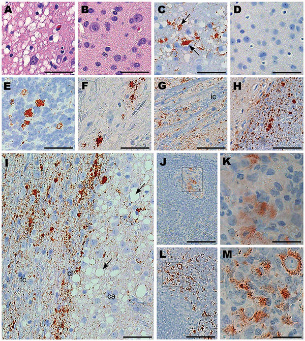
Figure 3. Immunohistochemical analysis of squirrel monkeys infected with chronic wasting disease (CWD) agent. Panels A, C, and E–M are from squirrel monkeys infected with CWD. Panels B and D are from an uninfected monkey showing no pathologic changes or positive staining for protease-resistant prion protein (PrPres). Panels A and B, cerebral cortex stained with hematoxylin and eosin; panels C and D, thalamus stained with antibody 3F4 against PrP (arrows); panels E and F, cerebellar granular cell layer and spinal cord, respectively, stained with antibody 3F4; panel G, gray matter within the internal capsule stained with antibody D13 against PrP; panel H, corpus callosum (right) stained with antibody D13 showing more intense staining than the adjacent cortex (left); panel I, frontal cortex (fc), claustrum (cl), and caudate (ca) stained with antibody 3F4 (abundant vacuoles in the putamen [arrows]); panels J–M, lymphatic tissue stained with antibody 3F4; panels J and K, PrPres staining in spleen of monkey 322; panels L and M, PrPres-positive mesenteric lymph node from orally infected monkey 301. Rectangles in panels J and L show areas enlarged in panels K and M, respectively. Antibodies D13 and 3F4 showed similar results for each monkey regarding the distribution, characteristics, and plaque size of PrPres. Scale bars: panels A–I, 50 μm; panel J, 100 μm; panels K and M, 25 μm; panel L, 250 μm.