Volume 17, Number 9—September 2011
Research
Classical Bovine Spongiform Encephalopathy by Transmission of H-Type Prion in Homologous Prion Protein Context
Juan-María Torres , Olivier Andréoletti, Caroline Lacroux, Irene Prieto, Patricia Lorenzo, Magdalena Larska, Thierry Baron, and Juan-Carlos Espinosa
Figure 4
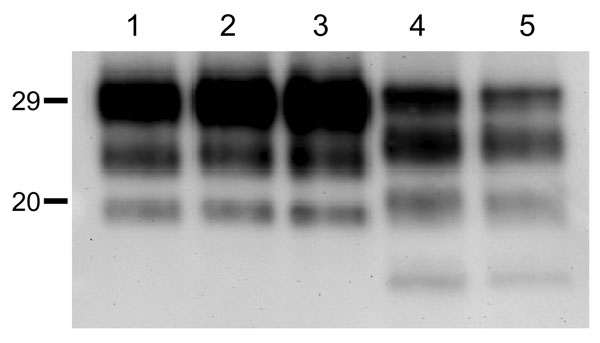
Figure 4. Western blot analyses of brain protease-resistant prion protein (PrPres) from BSE-H infected mice by using Saf84 monoclonal antibody. Tg110 mice infected with isolate 02-2695 (lanes 2 and 3) or 45 (lane 4) at first passage showing either high-type (lane 2) or classical BSE–like PrPres molecular profile (lanes 3 and 4). The BSE-H isolate (02–2695) (lane 1) and a BSE-C isolate (lane 5) were included for comparison. Similar quantities of PrPres were loaded in each lane. Values to the left indicate molecular mass in kDa. BSE, bovine spongiform encephalopathy; BSE-H, unglycosylated PrPres that is higher than BSE-C; BSE-C, classical BSE.
Page created: September 06, 2011
Page updated: September 06, 2011
Page reviewed: September 06, 2011
The conclusions, findings, and opinions expressed by authors contributing to this journal do not necessarily reflect the official position of the U.S. Department of Health and Human Services, the Public Health Service, the Centers for Disease Control and Prevention, or the authors' affiliated institutions. Use of trade names is for identification only and does not imply endorsement by any of the groups named above.