Skip directly to site content Skip directly to page options Skip directly to A-Z link Skip directly to A-Z link Skip directly to A-Z link
Volume 17, Number 9—September 2011
Research

Classical Bovine Spongiform Encephalopathy by Transmission of H-Type Prion in Homologous Prion Protein Context

Juan-María TorresComments to Author , Olivier Andréoletti, Caroline Lacroux, Irene Prieto, Patricia Lorenzo, Magdalena Larska, Thierry Baron, and Juan-Carlos Espinosa
Author affiliations: Author affiliations: Centro de Investigación en Sanidad Animal, Madrid, Spain (J.-M. Torres, I. Prieto, P. Lorenzo, M. Larska, J.-C. Espinosa); Ecole Nationale Vétérinaire de Toulouse, Toulouse, France (O. Andréoletti, C. Lacroux); Agence Francaise de Sécurité Sanitaire des Aliments, Lyon, France (T. Baron)

Main Article

Figure 7

Abnormal isoform of host-encoded prion protein (PrPSc) deposition patterns in brain and spleen from Tg110 mice infected with BSE-H. A–C) Paraffin-embedded tissue (PET) blots of representative coronal sections at the level of the hippocampus from Tg110 mice infected with atypical BSE-H (isolate 02-2695, first passage) showing either high-type (A) or classical-type protease-resistant prion protein (PrPres) phenotype (B). PET blot from Tg110 mice infected with BSE-C (C) is included for comparison.

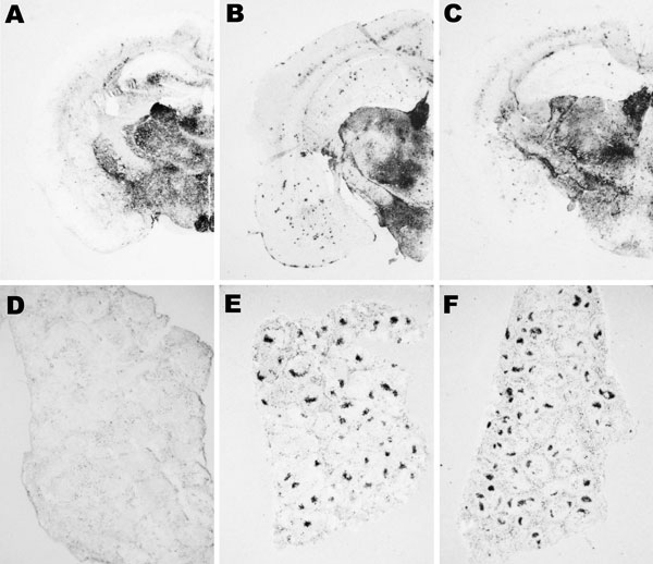
Figure 7. Abnormal isoform of host-encoded prion protein (PrPSc) deposition patterns in brain and spleen from Tg110 mice infected with BSE-H. A–C) Paraffin-embedded tissue (PET) blots of representative coronal sections at the level of the hippocampus from Tg110 mice infected with atypical BSE-H (isolate 02-2695, first passage) showing either high-type (A) or classical-type protease-resistant prion protein (PrPres) phenotype (B). PET blot from Tg110 mice infected with BSE-C (C) is included for comparison. D–F) PET blots of representative spleen sections: Tg110 mice infected with atypical BSE-H (isolate 02-2695) showing high-type PrPres in the brain were consistently scored as negative for PrPSc detection (D), whereas clear PrPSc deposits were always detected in BSE-H infected mice exhibiting BSE-C–like features (E), as in mice infected with BSE-C (F). Original magnification levels: panels A–C, ×20; panels D–F, ×6.BSE, bovine spongiform encephalopathy; BSE-H, unglycosylated PrPres that is higher than BSE-C; BSE-C, classical BSE.

Main Article

Page created: September 06, 2011
Page updated: September 06, 2011
Page reviewed: September 06, 2011
The conclusions, findings, and opinions expressed by authors contributing to this journal do not necessarily reflect the official position of the U.S. Department of Health and Human Services, the Public Health Service, the Centers for Disease Control and Prevention, or the authors' affiliated institutions. Use of trade names is for identification only and does not imply endorsement by any of the groups named above.
file_external