Volume 20, Number 5—May 2014
Research
Bovine Leukemia Virus DNA in Human Breast Tissue
Figure 1
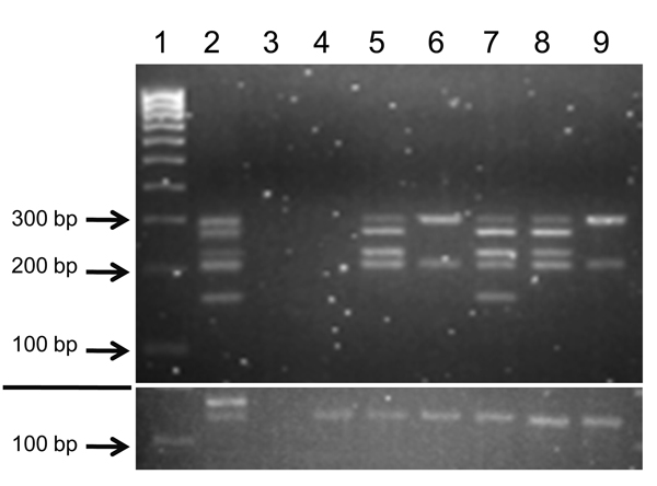
Figure 1. Amplification of bovine leukemia virus (BLV) genome regions in human breast tissue specimensNested liquid-phase PCR, using primers from 5 BLV genome regions, was used to amplify products from DNA extracted from breast tissues of 6 human donorsPCR products for each tissue were loaded into 1 well and separated by agarose gel (3.5%) electrophoresis on the basis of size differences: long terminal repeat, 290 bp; group-specific antigen, 272 bp; envelope, 230 bp; trans-activating gene of the X region, 206 bp; polymerase, 157 bpThe section below the white line shows the glyceraldehyde 3-phosphate dehydrogenase amplification of each sample as an indicator of DNA qualityLane 1, molecular weight marker (HyperLadder IV; Bioline, Taunton, MA, USA); lane 2, fetal lamb kidney cell line, positive control; lane 3, no-template-DNA negative control (water substituted for DNA template); lane 4, human sample 143; lane 5, human sample 236; lane 6, human sample 010; lane 7, human sample 20874; lane 8, human sample 23803; lane 9, human sample 0253.
1Current affiliation: Texas A&M Health Science Center College of Medicine, College Station, Texas, USA.