Volume 23, Number 3—March 2017
Research
Zika Virus RNA Replication and Persistence in Brain and Placental Tissue
Figure 3
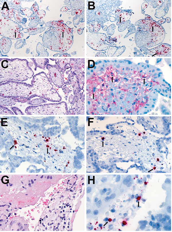
Figure 3. Localization of Zika virus RNA by ISH in placental tissues of women after spontaneous abortion. A) ISH with use of antisense probe. Zika virus genomic RNA localization in placental chorionic villi, predominantly within Hofbauer cells (red stain, arrows), of a case-patient who had spontaneous abortion at 11 wk gestation (case-patient no. 56). Original magnification ×10). B) ISH with use of sense probe. Serial section showing negative–strand replicative RNA intermediates (red stain, arrows) in the same cells shown in panel A. Original magnification ×10. C) Hematoxylin and eosin stain of placental tissue of a case-patient who experienced spontaneous abortion at 8 wk gestation (case-patient no. 47). Original magnification ×20. D) Immunostaining for CD163 highlighting villous Hofbauer cells in a serial section as seen in panel C. Original magnification ×63. E) ISH with use of antisense probe. Zika virus genomic RNA as seen in a serial section from the same case-patient as in panel C, showing staining within Hofbauer cells (red stain, arrows) of placental chorionic villi. Original magnification ×40. F) ISH with use of sense probe. Serial section showing negative-strand replicative RNA intermediates (red stain, arrows) in the same cells as shown in panel E. Original magnification ×40. G) Hematoxylin and eosin stain from the same case-patient as in panel C, showing inflammatory cell infiltrates in maternal side of placenta. Original magnification ×63. H) ISH with use of sense probe. Negative-strand replicative RNA intermediates (red stain, arrows) in inflammatory cells in a serial section. Original magnification ×63. ISH, in situ hybridization.