Volume 23, Number 8—August 2017
Research
Characterization of Fitzroy River Virus and Serologic Evidence of Human and Animal Infection
Cheryl A. Johansen1 , Simon H. Williams1, Lorna Melville, Jay Nicholson, Roy A. Hall, Helle Bielefeldt-Ohmann, Natalie A. Prow, Glenys R. Chidlow, Shani Wong, Rohini Sinha, David T. Williams, W. Ian Lipkin, and David W. Smith
Figure 3
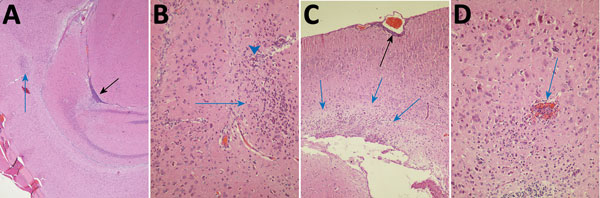
Figure 3. Photomicrographs of Fitzroy River virus (FRV)–induced meningoencephalitis in weanling mice inoculated with 1,000 infectious units of FRV. Panels show multifocal mild to severe perivascular and neuropil infiltration of lymphocytes and monocytes (blue arrows in A–C); meningitis in a sulcus (black arrow in A); glial cell activation with notable astrocytosis, neuron degeneration, and neuronophagia (arrowhead in B); occasional hemorrhage (blue arrow in D); mild periventricular spongiosis (blue arrows in C); and meningitis (black arrow in C). Hematoxylin and eosin staining. Original magnifications: A) ×40, B) ×400, C) ×100, D) ×400.
1These authors contributed equally to this article.
Page created: July 17, 2017
Page updated: July 17, 2017
Page reviewed: July 17, 2017
The conclusions, findings, and opinions expressed by authors contributing to this journal do not necessarily reflect the official position of the U.S. Department of Health and Human Services, the Public Health Service, the Centers for Disease Control and Prevention, or the authors' affiliated institutions. Use of trade names is for identification only and does not imply endorsement by any of the groups named above.