Skip directly to site content Skip directly to page options Skip directly to A-Z link Skip directly to A-Z link Skip directly to A-Z link
Volume 25, Number 1—January 2019
Research

Variable Protease-Sensitive Prionopathy Transmission to Bank Voles

Romolo Nonno1, Silvio Notari1, Michele Angelo Di Bari, Ignazio Cali, Laura Pirisinu, Claudia d’Agostino, Laura Cracco, Diane Kofskey, Ilaria Vanni, Jody Lavrich, Piero Parchi, Umberto Agrimi, and Pierluigi GambettiComments to Author 
Author affiliations: Istituto Superiore di Sanità, Rome, Italy (R. Nonno, M.A. Di Bari, L. Pirisinu, C. d’Agostino, I. Vanni, U. Agrimi); Case Western Reserve University, Cleveland, Ohio, USA (S. Notari, I. Cali, L. Cracco, D. Kofskey, J. Lavrich, P. Gambetti); University of Bologna, Bologna, Italy (P. Parchi); Istituto delle Scienze Neurologiche di Bologna, Bologna (P. Parchi)

Main Article

Figure 3

Representative paraffin-embedded tissue (PET) blots of protease-resistant, disease-related prion protein (resPrPD) distribution in phenotypes T1–T3 and controls. A) For T1, PrPD predominated in cerebral cortex (C), thalamus (T), superior colliculus (SC), lateral geniculate nucleus (LG), and substantia nigra (SN). A similar PrPD distribution was observed with transmission of sCJDMV1 used as type control. T2 showed a more uniform distribution in cerebral cortex and subcortical nuclei of an apparen

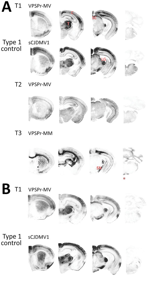
Figure 3. Representative paraffin-embedded tissue (PET) blots of protease-resistant, disease-related prion protein (resPrPD) distribution in phenotypes T1–T3 and controls. A) For T1, PrPD predominated in cerebral cortex (C), thalamus (T), superior colliculus (SC), lateral geniculate nucleus (LG), and substantia nigra (SN). A similar PrPD distribution was observed with transmission of sCJDMV1 used as type control. T2 showed a more uniform distribution in cerebral cortex and subcortical nuclei of an apparently lesser amount of PrPD; T3 appeared to preferentially affect the hemispheric white matter and other subcortical regions such as the alveus, the corpus callosum, the anterior commissure, and fascicles surrounding thalamus as well as other white matter formations such as fimbria, brachium of superior colliculus, medial lemniscus, and cerebral peduncles. Small amounts of PrPD were also observed in cerebellar and medullary white matter (asterisk [*]). B) PrPD T1 distribution resembled that of bank voles (bv) 109I after transmission of the same VPSPr-MV brain homogenate (compare with T1 in A). A similar distribution was also observed after inoculation with sCJDMV1. Left to right: coronal sections of telencephalon midlevel caudate nucleus; diencephalon midlevel thalamus; midbrain; and hindbrain-level medulla and cerebellum. sCJD, sporadic Creutzfeldt-Jakob disease; VPSPr, variably protease sensitive prionopathy.

Main Article

1These authors contributed equally to this article.

Page created: December 18, 2018
Page updated: December 18, 2018
Page reviewed: December 18, 2018
The conclusions, findings, and opinions expressed by authors contributing to this journal do not necessarily reflect the official position of the U.S. Department of Health and Human Services, the Public Health Service, the Centers for Disease Control and Prevention, or the authors' affiliated institutions. Use of trade names is for identification only and does not imply endorsement by any of the groups named above.
file_external