Volume 25, Number 6—June 2019
Research
Use of Single-Injection Recombinant Vesicular Stomatitis Virus Vaccine to Protect Nonhuman Primates Against Lethal Nipah Virus Disease
Figure 3
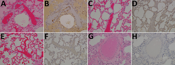
Figure 3. Results of testing for Nipah virus (NiV) in lung tissue from representative vaccinated African green monkeys (Chlorocebus aethiops). A, C, E, G) Hematoxylin and eosin staining; B, D, F, H) immunohistochemistry of tissues labeled with NiV N protein–specific polyclonal rabbit antibody. In stained tissue from the control animal (A), diffuse thickening of alveolar septae by moderate numbers of lymphocytes, plasma cells, polymerized fibrin, and edema fluid within the alveolar spaces were found; stained sections examined from the NiV F (C), NiV G (E), and NiV F/G (G) groups were unremarkable in comparison with sections from the control animal. In antibody-labeled tissue from the control animal (B), strong immunolabeling for NiV antigen with alveolar septae, scattered alveolar macrophages, and the endothelium of small caliber vessels were found, including syncytial cells with strong cytoplasmic immunolabeling for NiV antigen; no immunolabeling for NiV antigen was identified from the NiV F (D), NiV G (F), and NiV F/G (H) groups. Original magnification ×20.
1Current affiliation: Mayo Clinic, Rochester, Minnesota, USA.