Skip directly to site content Skip directly to page options Skip directly to A-Z link Skip directly to A-Z link Skip directly to A-Z link
Volume 25, Number 6—June 2019
Research

Use of Single-Injection Recombinant Vesicular Stomatitis Virus Vaccine to Protect Nonhuman Primates Against Lethal Nipah Virus Disease

Chad E. Mire, Joan B. Geisbert, Krystle N. Agans, Krista M. Versteeg, Daniel J. Deer, Benjamin A. Satterfield1, Karla A. Fenton, and Thomas W. GeisbertComments to Author 
Author affiliations: Galveston National Laboratory, Galveston, Texas, USA; University of Texas Medical Branch, Galveston

Main Article

Figure 4

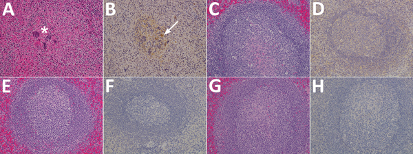
Results of testing for Nipah virus (NiV) in spleen tissue from representative vaccinated African green monkeys (Chlorocebus aethiops). A, C, E, G) Hematoxylin and eosin staining; B, D, F, H) immunohistochemistry of tissues labeled with NiV N protein–specific polyclonal rabbit antibody. In stained tissue from the control animal (A), moderate necrosis and drop of out the white pulp (*), with hemorrhage, and fibrin within germinal centers are seen; stained sections examined from the NiV F (C), NiV

Figure 4. Results of testing for Nipah virus (NiV) in spleen tissue from representative vaccinated African green monkeys (Chlorocebus aethiops). A, C, E, G) Hematoxylin and eosin staining; B, D, F, H) immunohistochemistry of tissues labeled with NiV N protein–specific polyclonal rabbit antibody. In stained tissue from the control animal (A), moderate necrosis and drop out of the white pulp (*), with hemorrhage, and fibrin within germinal centers are seen; stained sections examined from the NiV F (C), NiV G (E), and NiV F/G (G) groups were devoid of any significant lesions compared with sections from the control animal. In antibody-labeled tissues from the control animal (B), strong immunolabeling for NiV antigen with scattered mononuclear cells (white arrow) and syncytial cells within germinal centers were found, and the endothelium of small caliber vessels had strong cytoplasmic immunolabeling for NiV antigen; no immunolabeling for NiV antigen was identified from the NiV F (D), NiV G (F), and NiV F/G (H) groups. Original magnification ×20.

Main Article

1Current affiliation: Mayo Clinic, Rochester, Minnesota, USA.

Page created: May 20, 2019
Page updated: May 20, 2019
Page reviewed: May 20, 2019
The conclusions, findings, and opinions expressed by authors contributing to this journal do not necessarily reflect the official position of the U.S. Department of Health and Human Services, the Public Health Service, the Centers for Disease Control and Prevention, or the authors' affiliated institutions. Use of trade names is for identification only and does not imply endorsement by any of the groups named above.
file_external