Volume 26, Number 1—January 2020
Synopsis
Candidatus Mycoplasma haemohominis in Human, Japan
Norimichi Hattori1 , Makoto Kuroda1, Harutaka Katano1, Takahiro Takuma1, Takayoshi Ito1, Nana Arai, Ryo Yanai, Tsuyoshi Sekizuka, Sho Ishii, Yoko Miura, Takahiro Tokunaga, Hiroyuki Watanabe, Norihiro Nomura, Junichi Eguchi, Hideki Hasegawa, Tsuyoshi Nakamaki, Takaji Wakita, and Yoshihito Niki
Figure 6
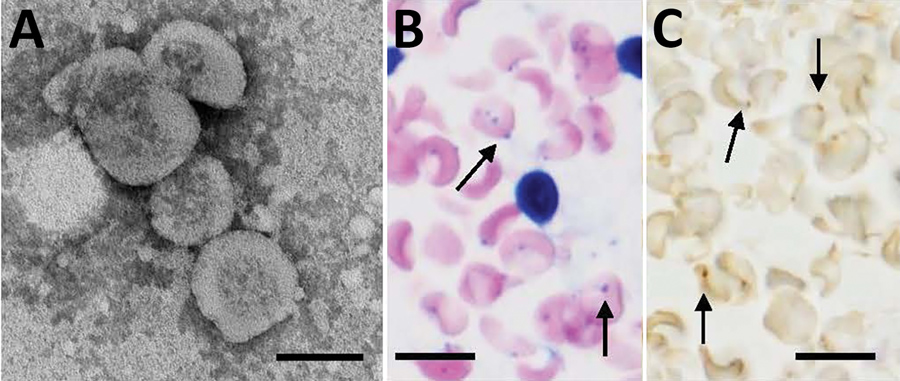
Figure 6. Morphologic features of Candidatus Mycoplasma haemohominis isolated from a serum sample of a 42-year-old man, Japan. A) Spheres are bacterial particles with a diameter of 300–600 nm. Negative stained; scale bar indicates 200 nm. B) Bacteria on the surface of erythrocytes (arrows in the left panel). In situ hybridization showing bacteria on the surface of erythrocytes (arrows in the right panel). Giemsa stained; scale bar indicates 10 μm.
1These authors contributed equally to this article.
Page created: December 18, 2019
Page updated: December 18, 2019
Page reviewed: December 18, 2019
The conclusions, findings, and opinions expressed by authors contributing to this journal do not necessarily reflect the official position of the U.S. Department of Health and Human Services, the Public Health Service, the Centers for Disease Control and Prevention, or the authors' affiliated institutions. Use of trade names is for identification only and does not imply endorsement by any of the groups named above.