Volume 26, Number 1—January 2020
Synopsis
Candidatus Mycoplasma haemohominis in Human, Japan
Norimichi Hattori1 , Makoto Kuroda1, Harutaka Katano1, Takahiro Takuma1, Takayoshi Ito1, Nana Arai, Ryo Yanai, Tsuyoshi Sekizuka, Sho Ishii, Yoko Miura, Takahiro Tokunaga, Hiroyuki Watanabe, Norihiro Nomura, Junichi Eguchi, Hideki Hasegawa, Tsuyoshi Nakamaki, Takaji Wakita, and Yoshihito Niki
Figure 3
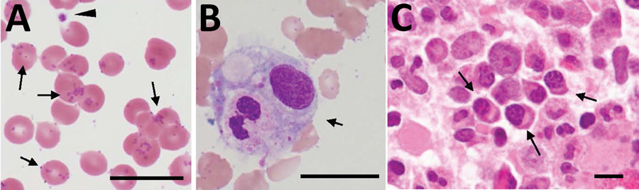
Figure 3. Distribution of Candidatus Mycoplasma haemohominis in a 42-year-old man, Japan. A) Peripheral blood smear showing coccoid forms. Small basophilic bodies are present on the surface of and outside erythrocytes (arrows). Arrowhead indicates a platelet. Giemsa stained. B) Hemophagocytosis (arrow) in bone marrow aspirate. Giemsa stained. C) Bone marrow biopsy specimen showing infiltration of plasma cells (arrows). Hematoxylin and eosin stained. Scale bars indicate 20 μm.
1These authors contributed equally to this article.
Page created: December 18, 2019
Page updated: December 18, 2019
Page reviewed: December 18, 2019
The conclusions, findings, and opinions expressed by authors contributing to this journal do not necessarily reflect the official position of the U.S. Department of Health and Human Services, the Public Health Service, the Centers for Disease Control and Prevention, or the authors' affiliated institutions. Use of trade names is for identification only and does not imply endorsement by any of the groups named above.