Disclaimer: Early release articles are not considered as final versions. Any changes will be reflected in the online version in the month the article is officially released.
Volume 32, Number 11—November 2026
Research
Emergence of Novel Macrolide-Resistant Mycoplasma pneumoniae Sequence Type 3 Subclades in Children after COVID-19 Pandemic, Taiwan

Figure 2
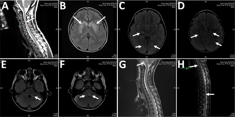
Figure 2. Magnetic resonance imaging (MRI) in selected patients with M. pneumoniae–related central nervous system complication in study of emergence of novel macrolide-resistant M. pneumoniae sequence type 3 subclades in children after COVID-19 pandemic, Taiwan, 2017–2024. A) Sagittal T2-weighted cervical spine MRI in patient 3, demonstrating a hyperintense lesion extending from C3 to C6 (arrow). B) Axial T2-FLAIR brain MRI of patient 5, showing a focal hyperintense lesion in the bilateral basal ganglia (arrows). C–F) Axial T2-FLAIR brain MRI scans of patient 10, revealing multiple hyperintense lesions involving the subcortical white matter of the bilateral parietal, temporal, and occipital lobes, as well as the midbrain, pons, and middle cerebellar peduncles, findings consistent with acute disseminated encephalomyelitis (arrows). G–H) Sagittal T2-weighted spine MRI of patient 10, indicating hyperintense lesions at the brainstem and at the T3 and T11 spinal levels (arrows).
1These senior authors contributed equally to this article.