Volume 32, Number 3—March 2026
Dispatch
Extraintestinal Entamoeba moshkovskii Infection, Eastern India
Sanjib Kumar Sardar, Ajanta Ghosal1, Tapas Haldar1, Basudev Ganguly, Koushik Das, and Sandipan Ganguly
Figure 1
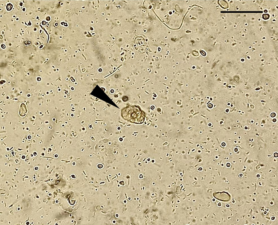
Figure 1. Microscopic view of Entamoeba moshkovskii trophozoites (arrowhead) observed in a pleural fluid sample recovered from a patient with an extraintestinal infection, eastern India. Slide used a wet-mount preparation,. Scale bar indicates 30 μm.
1These authors contributed equally to this article.
Page created: February 20, 2026
Page updated: March 20, 2026
Page reviewed: March 20, 2026
The conclusions, findings, and opinions expressed by authors contributing to this journal do not necessarily reflect the official position of the U.S. Department of Health and Human Services, the Public Health Service, the Centers for Disease Control and Prevention, or the authors' affiliated institutions. Use of trade names is for identification only and does not imply endorsement by any of the groups named above.