Volume 32, Number 3—March 2026
Research Letter
Mycobacterium riyadhense Pulmonary Disease after Relocation from Saudi Arabia, Japan
Takuya Ozawa, Takeshi Komine, Sohei Nakayama, Yusuke Suzuki, Naoki Hasegawa, Koichi Fukunaga, Ho Namkoong, Hanako Fukano, and Takanori Asakura
Figure 1
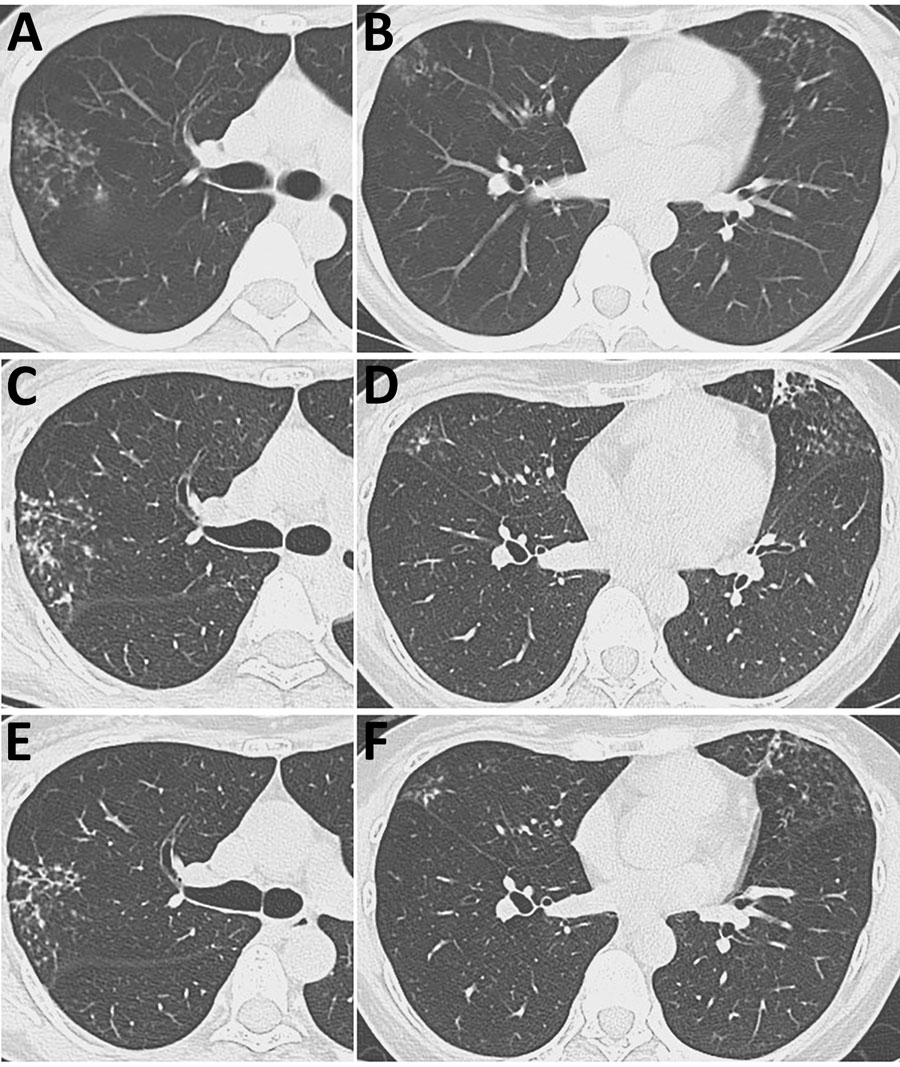
Figure 1. Serial axial chest computed tomography images over time from patient with Mycobacterium riyadhense pulmonary disease after relocation from Saudi Arabia, Japan. A, B) Images taken at initial hospital visit, demonstrating multiple scattered small nodular opacities in the right upper and middle lobes (A) and the lingular segment (B), accompanied by bronchial wall thickening. C, D) Images taken 2 years later, showing progression of the lesions in the right upper/middle lobes (C) and lingular segment (D). E, F) Images taken after treatment showing improvement of the lesions in the right upper/middle lobes (E) and lingular segment (F).
Page created: February 20, 2026
Page updated: March 20, 2026
Page reviewed: March 20, 2026
The conclusions, findings, and opinions expressed by authors contributing to this journal do not necessarily reflect the official position of the U.S. Department of Health and Human Services, the Public Health Service, the Centers for Disease Control and Prevention, or the authors' affiliated institutions. Use of trade names is for identification only and does not imply endorsement by any of the groups named above.