Volume 32, Number 3—March 2026
Research Letter
Mycobacterium riyadhense Pulmonary Disease after Relocation from Saudi Arabia, Japan
Cite This Article
Citation for Media
Abstract
We report a case of Mycobacterium riyadhense pulmonary disease in a patient who relocated from Saudi Arabia to Japan. Epidemiologic data and whole-genome analyses of the isolated strains suggested that the infection might have been acquired in Saudi Arabia and persisted, rather than a recent local acquisition in Japan.
Mycobacterium riyadhense, first isolated in Saudi Arabia, has been reported mainly in the Middle East (1) and sporadically elsewhere (2,3). We describe a patient who experienced slowly progressive pulmonary deterioration caused by M. riyadhense infection after she relocated from Saudi Arabia to Japan. Because M. riyadhense has not been reported in Japan, genomic analysis of the patient’s isolates was more consistent with within-host persistence of a preexisting infection than recent local acquisition from environmental exposure in Japan.
A 47-year-old woman was referred to Kitasato University Kitasato Institute Hospital (Tokyo, Japan) after granular opacities were detected in the right lung on screening. She had lived in Saudi Arabia for 2 years, where she had chronic exposure to sand and dust. A visibly contaminated, uncleaned air-conditioning unit at her home housed a bird’s nest for 7 months and remained in use. She took only showers and rarely cleaned the shower room. She also gardened regularly. Shortly before her initial visit for care, she returned to Japan, bringing back only clothing and no other household belongings. She resumed tub bathing; the showerhead was replaced 4 years after her return, while her illness was being monitored.
Figure 1

Figure 1. Serial axial chest computed tomography images over time from patient with Mycobacterium riyadhensepulmonary disease after relocation from Saudi Arabia, Japan. A, B) Images taken at initial hospital visit,...
Computed tomography (CT) revealed multiple small nodular opacities in the right upper and middle lobes and the lingular segment, along with bronchial wall thickening; those findings suggested the nodular bronchiectatic form of nontuberculous mycobacterial pulmonary disease (Figure 1, panel A, B). Bronchial wash from the right upper lobe was negative for acid-fast bacilli (AFB) by smear and culture. Because she was asymptomatic, we monitored her for 2 years. CT imaging showed progressive worsening (Figure 1, panel C, D). A repeat bronchial wash from the same site in the right upper lobe was negative by AFB smear; culture yielded M. riyadhense, identified by matrix-assisted laser desorption/ionization time-of-flight (MALDI-TOF) mass spectrometry using the MALDI Biotyper system with the Mycobacteria Library version 6.0 (Bruker, https://www.bruker.com) (4). Because she was asymptomatic without lung cavities, we deferred treatment.
Figure 2
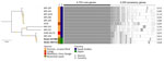
Figure 2. Midpoint-rooted maximum-likelihood tree based on 4,753 core genes of Mycobacterium riyadhense isolates from study of Mycobacterium riyadhensepulmonary disease after relocation from Saudi Arabia to Japan. Strains...
Five years after her initial visit, radiology-detected progression prompted a third bronchoscopy. Bronchial washes from 2 sites yielded M. riyadhense (strains 484719 and 537489), which we confirmed by MALDI-TOF mass spectrometry. We assembled draft genomes of the 2 strains from Illumina MiniSeq short-read sequencing data (https://www.illumina.com) using SPAdes version 3.15.5 (https://github.com/ablab/spades) (Appendix 1). Average nucleotide identity heatmap analysis using PyANI version 0.2.12 (https://github.com/widdowquinn/pyani) demonstrated that the isolates clustered with M. riyadhense, with >99.08% identity (Appendix 1 Figure 1; Appendix 2 Table 1). Phylogenetic analysis based on 4,753 core genes from 12 M. riyadhense genomes, including publicly available genomes from the National Center for Biotechnology Information database (Appendix), further showed that isolates from both specimens were closely related to strains reported from Saudi Arabia (Figure 2). We called 7 single-nucleotide polymorphisms (SNPs) using Snippy version 4.6.0 (https://github.com/tseemann/snippy) and Gubbins version 3.4 (https://github.com/nickjcroucher/gubbins) within the 2 isolated strains (Appendix 1 Figures 2, 3). Fourteen-day broth microdilution susceptibility testing showed favorable results (Appendix 2 Table 2). Four months later, sputum culture also yielded M. riyadhense. Azithromycin (250 mg/d) plus ethambutol (500 mg/d) achieved sputum culture conversion and radiologic improvement (Figure 1, panel E, F). Sputum cultures have remained negative on repeated follow-up.
We did not identify published case reports of M. riyadhense in Japan (Appendix). Recent studies showed that shower aerosols and certain soil types are common sources of NTM exposure (5,6). The patient had prolonged exposure to such environmental conditions while living in Saudi Arabia. Although the environmental reservoir of M. riyadhense is not completely defined, culture-independent surveys have detected M. riyadhense–consistent signatures in freshwater and soil samples, which suggests those habitats could represent potential sources of exposure (7,8). Our isolates differed from MR-193 by 11–12 SNPs, whereas they were substantially more distant from other publicly available genomes. However, neither a molecular clock nor SNP threshold for M. riyadhense has been established, so interpretation is limited; more genomes from the same cluster are needed to infer transmission. Nevertheless, considering the patient’s exposure history, clinical course, and the absence of previous detection reports of M. riyadhense in Japan, we considered within-host persistence of a preexisting infection to be a plausible explanation in this case.
No standard regimen for M. riyadhense infection has been established. Therapeutic approaches in previous cases have varied (1). A study summarizing previous cases of M. riyadhense (9) demonstrated efficacy of macrolide-based regimens combined with rifampin or fluoroquinolone, reporting a cure or improvement rate of 87.5%. In the case we describe, the isolate was susceptible to macrolides and other major drugs; therefore, we selected combination therapy with azithromycin and ethambutol. After initiating therapy, sputum cultures converted to negative within 2 months, with no evidence of recurrence. Subsequent imaging confirmed improvement in the lungs, providing further support for the efficacy of macrolide-based therapy against M. riyadhense.
Our findings contribute to understanding of the epidemiology and clinical course of M. riyadhense pulmonary disease. Given our whole-genome sequencing results and the absence of previous reports in Japan, this case might represent within-host persistence of a preexisting infection, distinct from recent local acquisition from environmental sources.
Dr. Ozawa is a physician specializing in respiratory medicine at the Division of Respiratory Medicine, Department of Internal Medicine, Keio University School of Medicine. He is pursuing a PhD, focusing on bronchiectasis and respiratory infectious diseases, particularly chronic infections such as nontuberculous mycobacterial pulmonary disease.
Acknowledgments
We thank Osamu Takeuchi and Masaharu Taga for their assistance with the transportation of mycobacteria.
This study was supported by a grant-in-aid for scientific research, Japan Society for the Promotion of Science (grant nos. JP25K02695 to T.A., JP22K16382 to H.F., JP24K19189 to T.K.).
Author contributions: T.O. and T.A. prepared the initial draft of the manuscript. S.N., Y.S., N.H., K.F., H.N., and H.F. revised subsequent versions. T.A. was responsible for the clinical management of the patient. T.K. and H.F. contributed to the bacteriologic diagnosis. All authors contributed to and approved the final manuscript.
References
- Varghese B, Enani MA, Althawadi S, Johani S, Fernandez GM, Al-Ghafli H, et al. Mycobacterium riyadhense in Saudi Arabia. Emerg Infect Dis. 2017;23:1732–4. DOIPubMedGoogle Scholar
- Godreuil S, Marchandin H, Michon AL, Ponsada M, Chyderiotis G, Brisou P, et al. Mycobacterium riyadhense pulmonary infection, France and Bahrain. Emerg Infect Dis. 2012;18:176–8. DOIPubMedGoogle Scholar
- Choi JI, Lim JH, Kim SR, Lee SH, Park JS, Seo KW, et al. Lung infection caused by Mycobacterium riyadhense confused with Mycobacterium tuberculosis: the first case in Korea. Ann Lab Med. 2012;32:298–303. DOIPubMedGoogle Scholar
- Markanović M, Makek MJ, Glodić G, Kuliš T, Mareković I. Evaluation and clinical impact of MALDI Biotyper Mycobacteria Library v6.0 for identification of nontuberculous mycobacteria by MALDI-TOF mass spectrometry. J Mass Spectrom. 2023;58:
e4915 . DOIPubMedGoogle Scholar - DeFlorio-Barker S, Egorov A, Smith GS, Murphy MS, Stout JE, Ghio AJ, et al. Environmental risk factors associated with pulmonary isolation of nontuberculous mycobacteria, a population-based study in the southeastern United States. Sci Total Environ. 2021;763:
144552 . DOIPubMedGoogle Scholar - Tzou CL, Dirac MA, Becker AL, Beck NK, Weigel KM, Meschke JS, et al. Association between Mycobacterium avium complex pulmonary disease and mycobacteria in home water and soil. Ann Am Thorac Soc. 2020;17:57–62. DOIPubMedGoogle Scholar
- King HC, Khera-Butler T, James P, Oakley BB, Erenso G, Aseffa A, et al. Environmental reservoirs of pathogenic mycobacteria across the Ethiopian biogeographical landscape. PLoS One. 2017;12:
e0173811 . DOIPubMedGoogle Scholar - Pontiroli A, Khera TT, Oakley BB, Mason S, Dowd SE, Travis ER, et al. Prospecting environmental mycobacteria: combined molecular approaches reveal unprecedented diversity. PLoS One. 2013;8:
e68648 . DOIPubMedGoogle Scholar - Alsaeed M, Alanazi K, Alhamdan A, Faqihi M, Alibrahim A, Alshehri S, et al. Exploring Mycobacterium riyadhense: epidemiology, clinical presentation, and treatment outcome. Open Forum Infect Dis. 2025;12:ofaf461. DOIGoogle Scholar
Figures
Cite This ArticleOriginal Publication Date: March 09, 2026
Table of Contents – Volume 32, Number 3—March 2026
| EID Search Options |
|---|
|
|
|
|
|
|
Please use the form below to submit correspondence to the authors or contact them at the following address:
Takanori Asakura, Division of Pulmonary Medicine, Department of Medicine, Keio University School of Medicine, 35 Shinanomachi, Shinjuku-ku, Tokyo 160-8582, Japan
Top