Skip directly to site content Skip directly to page options Skip directly to A-Z link Skip directly to A-Z link Skip directly to A-Z link

Disclaimer: Early release articles are not considered as final versions. Any changes will be reflected in the online version in the month the article is officially released.

Volume 32, Number 8—August 2026

Research

Detection of Highly Pathogenic Avian Influenza A(H5N1) Clade 2.3.4.4b Genotype D1.2 Virus in Swine after Experimental Inoculation

Hannah Seger, Amy L. Baker, Alexandra C. Buckley, Tavis K. Anderson, Alexey Markin, Alessandra Campos, Bruno Caetano Trindade, Marissa Vincent, Giovana Ciacci Zanella, Mia Torchetti, Kristina Lantz, and Bailey ArrudaComments to Author 
Author affiliation: Oak Ridge Institute for Science and Education, Oak Ridge, Tennessee, USA (H. Seger); US Department of Agriculture Agricultural Research Service National Animal Disease Center, Ames, Iowa, USA (H. Seger, A.L. Baker, A.C. Buckley, T.K. Anderson, A. Markin, A. Campos, B.C. Trindade, M. Vincent, G.C. Zanella, B. Arruda); Iowa State University College of Veterinary Medicine, Ames (G.C. Zanella); US Department of Agriculture Animal and Plant Health Inspection Service National Veterinary Services Laboratories, Ames (M. Torchetti, K. Lantz)

Main Article

Figure 1

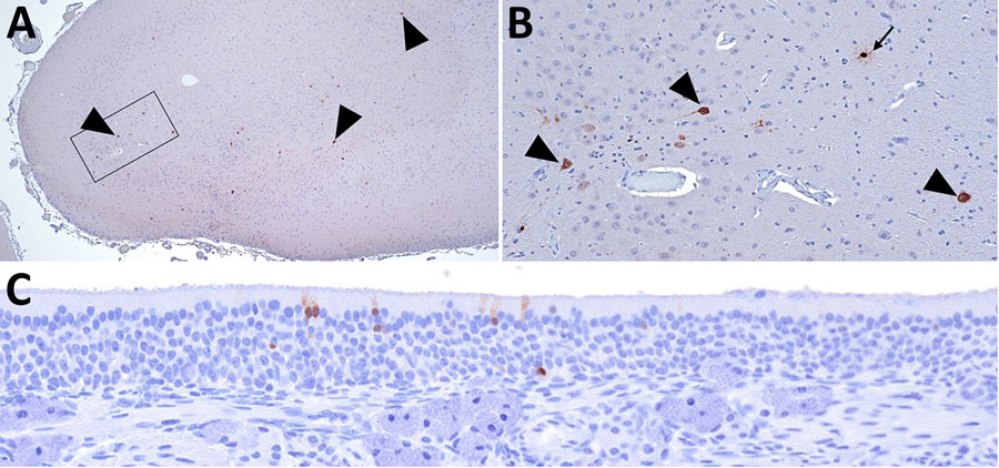
Immunohistochemical stains showing influenza A virus nucleoprotein (NP) antigen detection in pigs at 35 days postinoculation in an area with gliosis in study of systemic distribution and protracted detection of highly pathogenic avian influenza A(H5N1) clade 2.3.4.4b genotype D1.2 virus in swine after experimental inoculation. A) NP antigen within multiple cell types in the cerebrum of animal no. 650 (arrowheads). Original magnification ×40. B) Closer view of boxed area in panel A showing NP antigen within neurons (arrowhead) and glial cell (arrow). Original magnification ×200. C) NP antigen in the cytoplasm and nucleus of the olfactory epithelium of animal no. 649. Original magnification ×200.

Figure 1. Immunohistochemical stains showing influenza A virus nucleoprotein (NP) antigen detection in pigs at 35 days postinoculation in an area with gliosis in study of systemic distribution and protracted detection of highly pathogenic avian influenza A(H5N1) clade 2.3.4.4b genotype D1.2 virus in swine after experimental inoculation. A) NP antigen within multiple cell types in the cerebrum of animal no. 650 (arrowheads). Original magnification ×40. B) Closer view of boxed area in panel A showing NP antigen within neurons (arrowhead) and glial cell (arrow). Original magnification ×200. C) NP antigen in the cytoplasm and nucleus of the olfactory epithelium of animal no. 649. Original magnification ×200.

Main Article

Page created: June 23, 2026
Page updated: July 15, 2026
Page reviewed: July 15, 2026
The conclusions, findings, and opinions expressed by authors contributing to this journal do not necessarily reflect the official position of the U.S. Department of Health and Human Services, the Public Health Service, the Centers for Disease Control and Prevention, or the authors' affiliated institutions. Use of trade names is for identification only and does not imply endorsement by any of the groups named above.
file_external