Volume 32, Number 8—August 2026
Research
Detection of Highly Pathogenic Avian Influenza A(H5N1) Clade 2.3.4.4b Genotype D1.2 Virus in Swine after Experimental Inoculation

Figure 2
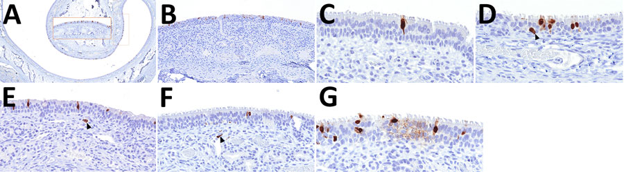
Figure 2. Distribution of influenza A virus nucleoprotein (NP) antigen in the nasal turbinates of pigs in study of systemic distribution and protracted detection of highly pathogenic avian influenza A(H5N1) clade 2.3.4.4b genotype D1.2 virus in swine after experimental inoculation. At 5 days postinoculation, NP antigen (brown) was commonly detected within the epithelial layer without (panel A; animal no. 648; original magnification ×40, inset ×200) and with (panel B; animal no. 642; original magnification ×200) notable loss of cilia of the turbinates, including the nucleus and cytoplasm of ciliated cells (panel C; animal no. 645; original magnification ×600) and less commonly nonciliated cells (panel D, arrowhead; animal no. 648; original magnification ×600). NP was also occasionally found within cells in the submucosa (panels E, F, arrowheads; animal nos. 641 and 648; original magnification ×600) and, rarely, at the cytoplasmic border of an aggregate of cells within the epithelium (panel G; animal no. 648; original magnification ×600).