Volume 32, Number 8—August 2026
Research
Detection of Highly Pathogenic Avian Influenza A(H5N1) Clade 2.3.4.4b Genotype D1.2 Virus in Swine after Experimental Inoculation

Figure 4
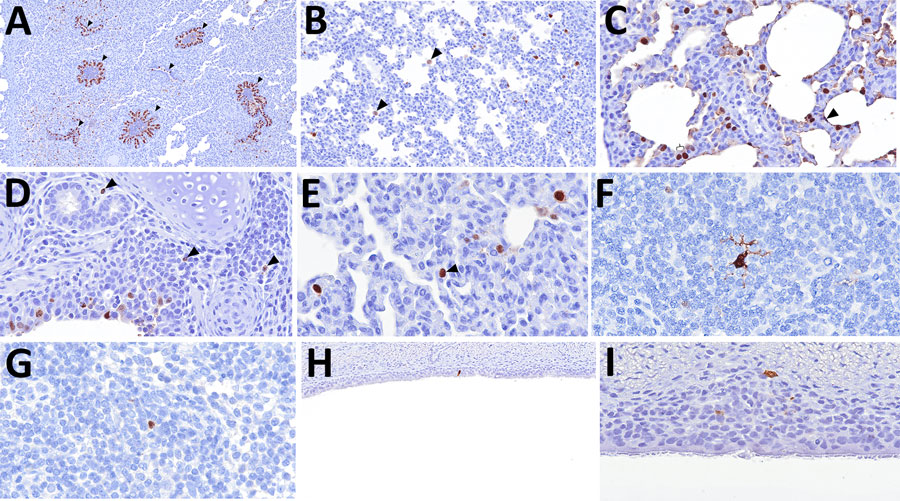
Figure 4. Distribution of influenza A virus nucleoprotein (NP) antigen in the lower respiratory tract and tracheobronchial lymph node of pigs in study of systemic distribution and protracted detection of highly pathogenic avian influenza A(H5N1) clade 2.3.4.4b genotype D1.2 virus in swine after experimental inoculation. At 5 days postinoculation, NP antigen (brown) was detected within the lower respiratory tract and associated lymphoid tissue. Immunolabeling in the lung was detected in the epithelium of conducting airways (panel A, arrowheads; animal no. 647; original magnification ×200), pulmonary alveolar macrophages (panel B; animal no. 645; original magnification ×200) lining the alveolar septa (arrowhead) and type II pneumocytes (brace), probable leukocytes within the bronchus-associated lymphoid tissue (panel D, arrowheads; animal no. 648; original magnification ×400), and leukocytes within the alveolar septa (panel E, arrowhead; animal no. 646; original magnification ×600). Viral antigen was also detected in the nucleus of leukocytes with morphologies consistent with an antigen-presenting leukocyte (panel F; animal no. 645; original magnification ×400) and lymphocyte (panel G; animal no. 646; original magnification ×600) in the tracheobronchial lymph node. NP antigen was only occasionally detected in the epithelial (panel H; animal no. 645; original magnification ×400) and submucosal (panel I; animal no. 643; original magnification ×600) layers of the trachea.