Volume 22, Number 8—August 2016
Research
Virulence and Evolution of West Nile Virus, Australia, 1960–2012
Natalie A. Prow12, Judith H. Edmonds2, David T. Williams, Yin X. Setoh, Helle Bielefeldt-Ohmann, Willy W. Suen, Jody Hobson-Peters, Andrew F. van den Hurk, Alyssa T. Pyke, Sonja Hall-Mendelin, Judith A. Northill, Cheryl A. Johansen, David Warrilow, Jianning Wang, Peter D. Kirkland, Stephen Doggett, Christy C. Andrade3, Aaron C. Brault, Alexander A. Khromykh4 , and Roy A. Hall4
Figure 2
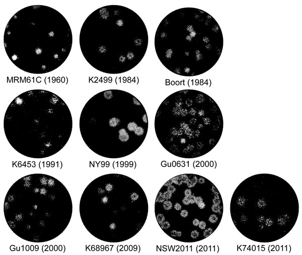
Figure 2. Plaque morphology of representative West Nile virus strains isolated in Australia, 1960–2012. Virus was allowed to adsorb to monolayers of Vero cells for 2 h at 37°C. The cells were then overlaid with Dulbecco Modified Eagle Medium containing 0.5% low melting point agarose and 2% fetal bovine serum. Four days after infection, the cells were fixed with 4% formaldehyde solution and stained with 0.2% crystal violet.
1Current affiliation: QIMR Berghofer Medical Research Institute, Herston, Queensland, Australia.
2These authors contributed equally to the major technical aspects of the work.
3Current affiliation: Willamette University, Salem, Oregon, USA.
4These authors served as joint senior authors.
Page created: July 15, 2016
Page updated: July 15, 2016
Page reviewed: July 15, 2016
The conclusions, findings, and opinions expressed by authors contributing to this journal do not necessarily reflect the official position of the U.S. Department of Health and Human Services, the Public Health Service, the Centers for Disease Control and Prevention, or the authors' affiliated institutions. Use of trade names is for identification only and does not imply endorsement by any of the groups named above.