Volume 23, Number 2—February 2017
Research
Swine Influenza Virus (H1N2) Characterization and Transmission in Ferrets, Chile
Nicolás Bravo-Vasquez1, Erik A. Karlsson1, Pedro Jimenez-Bluhm, Victoria Meliopoulos, Bryan Kaplan, Shauna Marvin, Valerie Cortez, Pamela Freiden, Melinda A. Beck, and Stacey Schultz-Cherry2
Figure 6
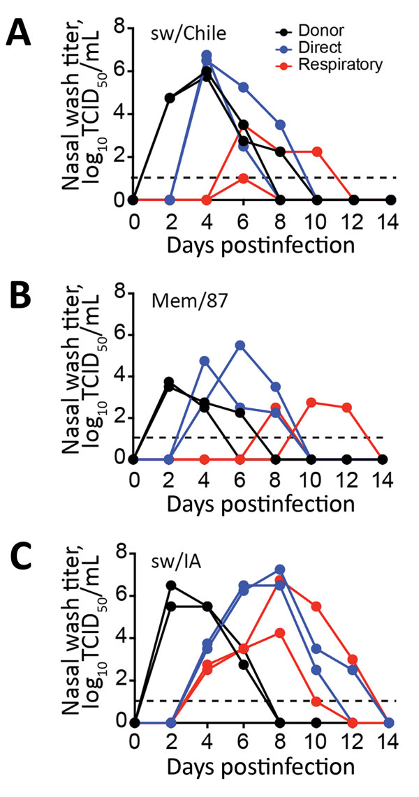
Figure 6. Evaluation of H1 virus transmission in ferrets. Donor ferrets (black lines; n = 2 ferrets/group) were inoculated with 106 50% tissue culture infectious dose (TCID50) units of H1 influenza virus sw/Chile (A), Mem/87 (B), or sw/IA (C) viruses. Naive ferrets (n = 2) were either placed in the same cage with the infected group (direct contact, blue lines) or housed in separate cages (respiratory transmission, red lines) and nasal washes were collected on the indicated day postinfection for virus quantification by TCID50 analysis. Lines represent individual animals. Dashed line represents limit of detection of the assay.
1These first authors contributed equally to this article.
2These senior authors contributed equally to this article.
Page created: January 17, 2017
Page updated: January 17, 2017
Page reviewed: January 17, 2017
The conclusions, findings, and opinions expressed by authors contributing to this journal do not necessarily reflect the official position of the U.S. Department of Health and Human Services, the Public Health Service, the Centers for Disease Control and Prevention, or the authors' affiliated institutions. Use of trade names is for identification only and does not imply endorsement by any of the groups named above.