Volume 25, Number 1—January 2019
Research
Association of Increased Receptor-Binding Avidity of Influenza A(H9N2) Viruses with Escape from Antibody-Based Immunity and Enhanced Zoonotic Potential
Figure 7

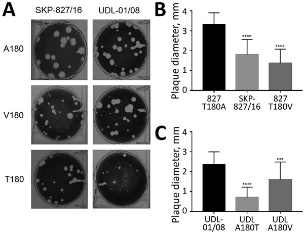
Figure 7. Plaque phenotype of influenza A(H9N2) viruses in UDL-01/08 and SKP-827/16 A/T/V180 variants in MDCK cells. A) Plaque morphology of wild-type UDL-01/08 (containing A180) and SKP-827/16 (containing T180) viruses and variants containing A/T/V180 substitutions. B, C) 30 plaques were selected for each virus, and ImageJ software (https://imagej.nih.gov/ij) was used to measure plaque diameter. Comparisons were conducted between viruses of the same hemagglutinin backbone with different substitutions (A/T/V180). Error bars indicate SEM. ***p<0.001; ****p<0.0001.
1Current affiliation: Imperial College London, London, UK.
2Current affiliation: Chinese Academy of Agricultural Sciences, Beijing, China.
Page created: December 18, 2018
Page updated: December 18, 2018
Page reviewed: December 18, 2018
The conclusions, findings, and opinions expressed by authors contributing to this journal do not necessarily reflect the official position of the U.S. Department of Health and Human Services, the Public Health Service, the Centers for Disease Control and Prevention, or the authors' affiliated institutions. Use of trade names is for identification only and does not imply endorsement by any of the groups named above.