Skip directly to site content Skip directly to page options Skip directly to A-Z link Skip directly to A-Z link Skip directly to A-Z link
Volume 30, Number 7—July 2024
Research

Sialic Acid Receptor Specificity in Mammary Gland of Dairy Cattle Infected with Highly Pathogenic Avian Influenza A(H5N1) Virus

Rahul K. Nelli1Comments to Author , Tyler A. Harm1, Chris Siepker1, Jennifer M. Groeltz-Thrush, Brianna Jones, Ning-Chieh Twu, Ariel S. Nenninger, Drew R. Magstadt, Eric R. Burrough, Pablo E. Piñeyro, Marta Mainenti, Silvia Carnaccini, Paul J. Plummer, and Todd M. BellComments to Author 
Author affiliations: Iowa State University College of Veterinary Medicine, Ames, Iowa, USA (R.K. Nelli, T.A. Harm, C. Siepker, J.M. Groeltz-Thrush, B. Jones, N.-C. Twu, A.S. Nenninger, D.R. Magstadt, E.R. Burrough, P.E. Piñeyro, M. Mainenti, P.J. Plummer, T.M. Bell); University of Georgia, Athens, Georgia, USA (S. Carnaccini)

Main Article

Figure 1

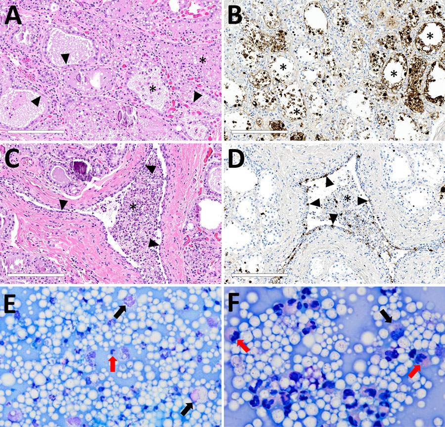
Microscopic mammary lesions of an index case in US dairy cattle naturally infected with highly pathogenic avian influenza A(H5N1) virus clade 2.3.4.4b. A) Mammary gland alveoli show epithelial attenuation and vacuolation (arrows), leading to degeneration with intraluminal sloughing and neutrophilic intraluminal inflammation (asterisk). Hematoxylin and eosin stain. B) Mammary glad alveoli show degenerative epithelial cells (asterisks) and strong intracytoplasmic and nuclear immunoreactivity to influenza A virus nucleoprotein. C) Cuboidal epithelium lining of the interlobular duct was markedly attenuated (arrowheads) with abundant intraluminal sloughing and neutrophilic inflammation (asterisk). Hematoxylin and eosin stain. D) Interlobular duct shows of attenuating and degenerative epithelium by intranuclear and cytoplasmic immunoreactivity (brown labeling) with immunopositive intraluminal debris and inflammation (asterisk). Scale bars indicate 200 μm. E, F) Modified Wright's stained representative cytology images of milk from a dairy cow with highly pathogenic avian influenza A(H5N1) virus infection, demonstrating moderate to marked neutrophilic inflammation (red arrows) with low numbers of vacuolated macrophages (black arrows) among large numbers of lipid vacuoles. A 100-cell count was performed, and nucleated cells were found to consist of 83% neutrophils, 12% macrophages, and 5% lymphocytes. Original magnifications ×500 for panel E and ×1,000 for panel F.

Figure 1. Microscopic mammary lesions of an index case in US dairy cattle naturally infected with highly pathogenic avian influenza A(H5N1) virus clade 2.3.4.4b. A) Mammary gland alveoli show epithelial attenuation and vacuolation (arrows), leading to degeneration with intraluminal sloughing and neutrophilic intraluminal inflammation (asterisk). Hematoxylin and eosin stain. B) Mammary glad alveoli show degenerative epithelial cells (asterisks) and strong intracytoplasmic and nuclear immunoreactivity to influenza A virus nucleoprotein. C) Cuboidal epithelium lining of the interlobular duct was markedly attenuated (arrowheads) with abundant intraluminal sloughing and neutrophilic inflammation (asterisk). Hematoxylin and eosin stain. D) Interlobular duct shows of attenuating and degenerative epithelium by intranuclear and cytoplasmic immunoreactivity (brown labeling) with immunopositive intraluminal debris and inflammation (asterisk). Scale bars indicate 200 μm. E, F) Modified Wright's stained representative cytology images of milk from a dairy cow with highly pathogenic avian influenza A(H5N1) virus infection, demonstrating moderate to marked neutrophilic inflammation (red arrows) with low numbers of vacuolated macrophages (black arrows) among large numbers of lipid vacuoles. A 100-cell count was performed, and nucleated cells were found to consist of 83% neutrophils, 12% macrophages, and 5% lymphocytes. Original magnifications ×500 for panel E and ×1,000 for panel F.

Main Article

1These first authors contributed equally to this article.

Page created: June 03, 2024
Page updated: June 22, 2024
Page reviewed: June 22, 2024
The conclusions, findings, and opinions expressed by authors contributing to this journal do not necessarily reflect the official position of the U.S. Department of Health and Human Services, the Public Health Service, the Centers for Disease Control and Prevention, or the authors' affiliated institutions. Use of trade names is for identification only and does not imply endorsement by any of the groups named above.
file_external